
CTBP2 Rabbit mAb, Unconjugated, Monoclonal
Artikelnummer:
ABB-A0463
- Bilder (8)


| Artikelname: | CTBP2 Rabbit mAb, Unconjugated, Monoclonal |
| Artikelnummer: | ABB-A0463 |
| Hersteller Artikelnummer: | A0463 |
| Alternativnummer: | ABB-A0463-20UL,ABB-A0463-100UL |
| Hersteller: | ABclonal |
| Wirt: | Rabbit |
| Kategorie: | Antikörper |
| Applikation: | ELISA, IF, IHC-P, IP, WB |
| Spezies Reaktivität: | Human |
| Immunogen: | Synthetic peptide. This information is considered to be commercially sensitive. |
| Konjugation: | Unconjugated |
| Alternative Synonym: | CTBP2, C-terminal-binding protein 2 |
| This gene produces alternative transcripts encoding two distinct proteins. One protein is a transcriptional repressor, while the other isoform is a major component of specialized synapses known as synaptic ribbons. Both proteins contain a NAD+ binding domain similar to NAD+-dependent 2-hydroxyacid dehydrogenases. A portion of the 3 untranslated region was used to map this gene to chromosome 21q21.3, however, it was noted that similar loci elsewhere in the genome are likely. Blast analysis shows that this gene is present on chromosome 10. Several transcript variants encoding two different isoforms have been found for this gene. |
| Application Verdünnung: | WB,1:500 - 1:1000|IHC-P,1:50 - 1:200|IF/ICC,1:50 - 1:200|IP,0.5µg-4µg antibody for 200µg-400µg extracts of whole cells|ELISA,Recommended starting concentration is 1 µg/mL. Please optimize the concentration based on your specific assay requirements. |
| Anwendungsbeschreibung: | Cross-Reactivity: Human,Mouse,Rat. ResearchArea: Epigenetics Nuclear Signaling,Cell Biology Developmental Biology,Neuroscience, Cell Type Marker,Neuron marker,Synapse marker. Shipping: Ice Bag |
|
|
Western blot analysis of various lysates using CTBP2 Rabbit mAb (A0463) at 1:1000 dilution. Secondary antibody: HRP-conjugated Goat anti-Rabbit IgG (H+L) (AS014) at 1:10000 dilution. Lysates/proteins: 25µg per lane. Blocking buffer: 3% nonfat dry milk in TBST. Detection: ECL Basic Kit (RM00020). Exposure time: 1s. |
|
|
Immunohistochemistry analysis of paraffin-embedded Human colon carcinoma using CTBP2 Rabbit mAb (A0463) at dilution of 1:100 (40x lens). High pressure antigen retrieval performed with 0.01M Citrate buffer (pH 6.0) prior to IHC staining. |
|
|
Immunohistochemistry analysis of paraffin-embedded Mouse kidney using CTBP2 Rabbit mAb (A0463) at dilution of 1:100 (40x lens). High pressure antigen retrieval performed with 0.01M Citrate buffer (pH 6.0) prior to IHC staining. |
|
|
Immunohistochemistry analysis of paraffin-embedded Rat ovary using CTBP2 Rabbit mAb (A0463) at dilution of 1:100 (40x lens). High pressure antigen retrieval performed with 0.01M Citrate buffer (pH 6.0) prior to IHC staining. |
|
|
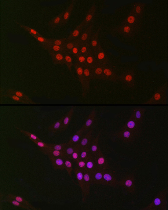
Immunofluorescence analysis of NIH/3T3 cells using CTBP2 Rabbit mAb (A0463) at dilution of 1:100 (40x lens). Secondary antibody: Cy3-conjugated Goat anti-Rabbit IgG (H+L) (AS007) at 1:500 dilution. Blue: DAPI for nuclear staining. |
|
|
Immunofluorescence analysis of PC-12 cells using CTBP2 Rabbit mAb (A0463) at dilution of 1:100 (40x lens). Secondary antibody: Cy3-conjugated Goat anti-Rabbit IgG (H+L) (AS007) at 1:500 dilution. Blue: DAPI for nuclear staining. |
|
|
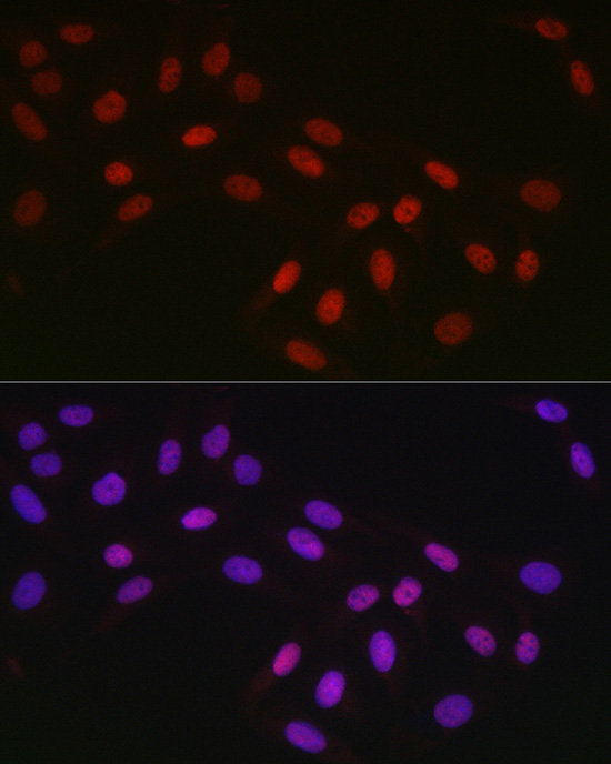
Immunofluorescence analysis of U2OS cells using CTBP2 Rabbit mAb (A0463) at dilution of 1:100 (40x lens). Secondary antibody: Cy3-conjugated Goat anti-Rabbit IgG (H+L) (AS007) at 1:500 dilution. Blue: DAPI for nuclear staining. |
|
|
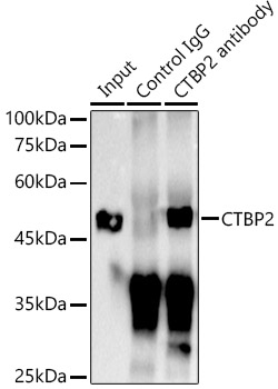
Immunoprecipitation analysis of 300 µg extracts of Hela cells using 3 µg CTBP2 antibody (A0463). Western blot was performed from the immunoprecipitate using CTBP2 (A0463) at a dilution of 1:1000. |
Produktgarantie und fachkundiger Support
