
MTAP Rabbit pAb, Unconjugated, Polyclonal
Artikelnummer:
ABB-A1049
- Bilder (8)


| Artikelname: | MTAP Rabbit pAb, Unconjugated, Polyclonal |
| Artikelnummer: | ABB-A1049 |
| Hersteller Artikelnummer: | A1049 |
| Alternativnummer: | ABB-A1049-100UL,ABB-A1049-20UL,ABB-A1049-500UL,ABB-A1049-1000UL |
| Hersteller: | ABclonal |
| Wirt: | Rabbit |
| Kategorie: | Antikörper |
| Applikation: | ELISA, IF, IHC-P, WB |
| Spezies Reaktivität: | Human |
| Immunogen: | Recombinant protein (or fragment).This information is considered to be commercially sensitive. |
| Konjugation: | Unconjugated |
| Alternative Synonym: | BDMF, MSAP, DMSFH, LGMBF, DMSMFH, c86fus, HEL-249, MTAP |
| This gene encodes an enzyme that plays a major role in polyamine metabolism and is important for the salvage pathway of both adenine and methionine. The encoded enzyme is deficient in many cancers. Multiple alternatively spliced transcript variants have been described for this gene. |
| Klonalität: | Polyclonal |
| Molekulargewicht: | 31kDa |
| NCBI: | 4507 |
| UniProt: | Q13126 |
| Reinheit: | Affinity purification |
| Sequenz: | MASGTTTTAVKIGIIGGTGLDDPEILEGRTEKYVDTPFGKPSDALILGKIKNVDCVLLARHGRQHTIMPSKVNYQANIWALKEEGCTHVIVTTACGSLREEIQPGDIVIIDQFIDRTTMRPQSFYDGSHSCARGVCHIPMAEPFCPKTREVLIETAKKLGLRCHSKGTMVTIEGPRFSSRAESFMFRTWGADVINMTTVPEVVLAKEAGICYASIAMATDYDCWKEHEEAVSVDRVLKTLKENANKAKSLLLTTI |
| Target-Kategorie: | MTAP |
| Antibody Type: | Primary Antibody |
| Application Verdünnung: | WB,1:500 - 1:1000|IHC-P,1:50 - 1:200|IF/ICC,1:50 - 1:200|ELISA,Recommended starting concentration is 1 µg/mL. Please optimize the concentration based on your specific assay requirements. |
| Anwendungsbeschreibung: | Cross-Reactivity: Human,Mouse,Rat. ResearchArea: Cancer,Signal Transduction,Endocrine Metabolism,Amino acid metabolism. Shipping: Ice Bag |
|
|
Western blot analysis of various lysates using MTAP Rabbit pAb (A1049) at 1:1000 dilution. Secondary antibody: HRP-conjugated Goat anti-Rabbit IgG (H+L) (AS014) at 1:10000 dilution. Lysates/proteins: 25µg per lane. Blocking buffer: 3% nonfat dry milk in TBST. Detection: ECL Basic Kit (RM00020). Exposure time: 180s. |
|
|
Immunohistochemistry analysis of paraffin-embedded Human colon carcinoma using MTAP Rabbit pAb (A1049) at dilution of 1:50 (40x lens). High pressure antigen retrieval performed with 0.01M Citrate buffer (pH 6.0) prior to IHC staining. |
|
|
Western blot analysis of lysates from NIH/3T3 cells, using MTAP Rabbit pAb (A1049) at 1:1000 dilution. Secondary antibody: HRP-conjugated Goat anti-Rabbit IgG (H+L) (AS014) at 1:10000 dilution. Lysates/proteins: 25µg per lane. Blocking buffer: 3% nonfat dry milk in TBST. Detection: ECL Basic Kit (RM00020). Exposure time: 30s. |
|
|
Immunohistochemistry analysis of paraffin-embedded Mouse kidney using MTAP Rabbit pAb (A1049) at dilution of 1:50 (40x lens). High pressure antigen retrieval performed with 0.01M Citrate buffer (pH 6.0) prior to IHC staining. |
|
|
Immunohistochemistry analysis of paraffin-embedded Rat ovary using MTAP Rabbit pAb (A1049) at dilution of 1:50 (40x lens). High pressure antigen retrieval performed with 0.01M Citrate buffer (pH 6.0) prior to IHC staining. |
|
|
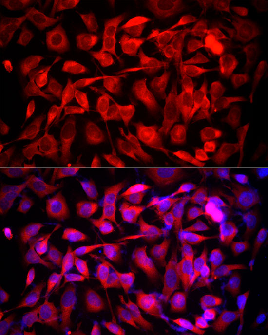
Immunofluorescence analysis of HeLa cells using MTAP Rabbit pAb (A1049) at dilution of 1:50 (40x lens). Secondary antibody: Cy3-conjugated Goat anti-Rabbit IgG (H+L) (AS007) at 1:500 dilution. Blue: DAPI for nuclear staining. |
|
|
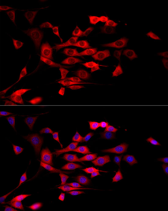
Immunofluorescence analysis of NIH/3T3 cells using MTAP Rabbit pAb (A1049) at dilution of 1:50 (40x lens). Secondary antibody: Cy3-conjugated Goat anti-Rabbit IgG (H+L) (AS007) at 1:500 dilution. Blue: DAPI for nuclear staining. |
|
|
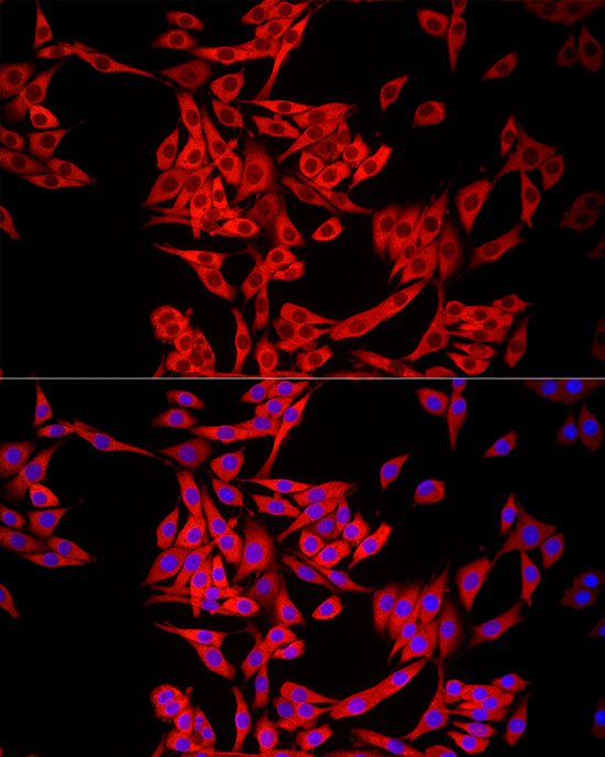
Immunofluorescence analysis of PC-12 cells using MTAP Rabbit pAb (A1049) at dilution of 1:50 (40x lens). Secondary antibody: Cy3-conjugated Goat anti-Rabbit IgG (H+L) (AS007) at 1:500 dilution. Blue: DAPI for nuclear staining. |
Produktgarantie und fachkundiger Support
