
TIMM50 Rabbit mAb, Unconjugated, Monoclonal
Artikelnummer:
ABB-A1536
- Bilder (8)


| Artikelname: | TIMM50 Rabbit mAb, Unconjugated, Monoclonal |
| Artikelnummer: | ABB-A1536 |
| Hersteller Artikelnummer: | A1536 |
| Alternativnummer: | ABB-A1536-100UL,ABB-A1536-20UL |
| Hersteller: | ABclonal |
| Wirt: | Rabbit |
| Kategorie: | Antikörper |
| Applikation: | ELISA, IF, IHC-P, WB |
| Spezies Reaktivität: | Human |
| Immunogen: | Synthetic peptide. This information is considered to be commercially sensitive. |
| Konjugation: | Unconjugated |
| Alternative Synonym: | MGCA9, TIM50, TIM50L, TIMM50 |
| This gene encodes a subunit of the TIM23 inner mitochondrial membrane translocase complex. The encoded protein functions as the receptor subunit that recognizes the mitochondrial targeting signal, or presequence, on protein cargo that is destined for the mitochondrial inner membrane and matrix. This protein may also play a role in maintaining the membrane permeability barrier, and knockdown of this gene in human cells results in the release of cytochrome c and apoptosis. |
| Application Verdünnung: | WB,1:500 - 1:1000|IF/ICC,1:50 - 1:200|IF-P,1:50 - 1:200|IHC-P,1:50 - 1:200|ELISA,Recommended starting concentration is 1 µg/mL. Please optimize the concentration based on your specific assay requirements. |
| Anwendungsbeschreibung: | Cross-Reactivity: Human,Mouse,Rat. ResearchArea: Endocrine Metabolism,Mitochondrial metabolism. Shipping: Ice Bag |
|
|
Western blot analysis of various lysates using TIMM50 Rabbit mAb (A1536) at 1:1000 dilution. Secondary antibody: HRP-conjugated Goat anti-Rabbit IgG (H+L) (AS014) at 1:10000 dilution. Lysates/proteins: 25µg per lane. Blocking buffer: 3% nonfat dry milk in TBST. Detection: ECL Basic Kit (RM00020). Exposure time: 30s. |
|
|
Western blot analysis of various lysates using TIMM50 Rabbit mAb (A1536) at 1:1000 dilution. Secondary antibody: HRP-conjugated Goat anti-Rabbit IgG (H+L) (AS014) at 1:10000 dilution. Lysates/proteins: 25µg per lane. Blocking buffer: 3% nonfat dry milk in TBST. Detection: ECL Basic Kit (RM00020). Exposure time: 60s. |
|
|
Immunohistochemistry analysis of paraffin-embeddedMouse kidney tissue usingTIMM50 Rabbit mAb(A1536) at a dilution of 1:200 (40x lens).High pressure antigen retrieval was performed with 0.01 M Tris-EDTA buffer (pH 9.0) prior to IHC staining. |
|
|
Immunohistochemistry analysis of paraffin-embeddedHuman liver tissue usingTIMM50 Rabbit mAb(A1536) at a dilution of 1:200 (40x lens).High pressure antigen retrieval was performed with 0.01 M Tris-EDTA buffer (pH 9.0) prior to IHC staining. |
|
|
Immunohistochemistry analysis of paraffin-embeddedRat kidney tissue usingTIMM50 Rabbit mAb(A1536) at a dilution of 1:200 (40x lens).High pressure antigen retrieval was performed with 0.01 M Tris-EDTA buffer (pH 9.0) prior to IHC staining. |
|
|
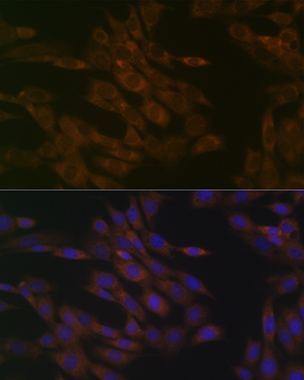
Confocal imaging of NIH/3T3 cells usingTIMM50 Rabbit mAb (A1536, dilution 1:100) followed by a further incubation with Cy3 Goat Anti-Rabbit IgG (H+L) (AS007, dilution 1:500) (Red). The cells were counterstained with alpha-Tubulin Mouse mAb (AC012, dilution 1:400) followed by incubation with ABflo 488-conjugated Goat Anti-Mouse IgG (H+L) Ab (AS076, dilution 1:500) (Green). DAPI was used for nuclear staining (Blue). Objective: 100x. |
|
|
Confocal imaging ofparaffin-embedded Mouse kidney tissue usingTIMM50 Rabbit mAb (A1536, dilution 1:100) followed by a further incubation with Cy3 Goat Anti-Rabbit IgG (H+L) (AS007, dilution 1:500) (Red). DAPI was used for nuclear staining (Blue). Objective: 40x. Perform high pressure antigen retrieval with 0.01 M citrate buffer (pH 6.0) prior to IF staining. |
|
|
Confocal imaging ofparaffin-embedded Human kidney tissue usingTIMM50 Rabbit mAb (A1536, dilution 1:100) followed by a further incubation with Cy3 Goat Anti-Rabbit IgG (H+L) (AS007, dilution 1:500) (Red). DAPI was used for nuclear staining (Blue). Objective: 40x. Perform high pressure antigen retrieval with 0.01 M citrate buffer (pH 6.0) prior to IF staining. |
Produktgarantie und fachkundiger Support
