
IL2 Rabbit pAb, Unconjugated, Polyclonal
Artikelnummer:
ABB-A16317
- Bilder (8)

| Artikelname: | IL2 Rabbit pAb, Unconjugated, Polyclonal |
| Artikelnummer: | ABB-A16317 |
| Hersteller Artikelnummer: | A16317 |
| Alternativnummer: | ABB-A16317-20UL,ABB-A16317-100UL,ABB-A16317-1000UL,ABB-A16317-500UL |
| Hersteller: | ABclonal |
| Wirt: | Rabbit |
| Kategorie: | Antikörper |
| Applikation: | ELISA, IHC-P, WB |
| Spezies Reaktivität: | Human |
| Immunogen: | Recombinant protein (or fragment).This information is considered to be commercially sensitive. |
| Konjugation: | Unconjugated |
| Alternative Synonym: | IL-2, TCGF, lymphokine, IL2 |
| This gene is a member of the interleukin 2 (IL2) cytokine subfamily which includes IL4, IL7, IL9, IL15, IL21, erythropoietin, and thrombopoietin. The protein encoded by this gene is a secreted cytokine produced by activated CD4+ and CD8+ T lymphocytes, that is important for the proliferation of T and B lymphocytes. The receptor of this cytokine (IL2R) is a heterotrimeric protein complex whose gamma chain is also shared by IL4 and IL7. The expression of this gene in mature thymocytes is monoallelic, which represents an unusual regulatory mode for controlling the precise expression of a single gene. The targeted disruption of a similar gene in mice leads to ulcerative colitis-like disease, which suggests an essential role of this gene in the immune response to antigenic stimuli. |
| Klonalität: | Polyclonal |
| Molekulargewicht: | 18kDa |
| NCBI: | 3558 |
| UniProt: | P60568 |
| Reinheit: | Affinity purification |
| Sequenz: | APTSSSTKKTQLQLEHLLLDLQMILNGINNYKNPKLTRMLTFKFYMPKKATELKHLQCLEEELKPLEEVLNLAQSKNFHLRPRDLISNINVIVLELKGSETTFMCEYADETATIVEFLNRWITFCQSIISTLT |
| Target-Kategorie: | IL2 |
| Antibody Type: | Primary Antibody |
| Application Verdünnung: | WB,1:500 - 1:1000|IHC-P,1:50 - 1:200|ELISA,Recommended starting concentration is 1 µg/mL. Please optimize the concentration based on your specific assay requirements. |
| Anwendungsbeschreibung: | Cross-Reactivity: Human,Mouse,Rat. ResearchArea: Cell Biology Developmental Biology,Apoptosis,Growth factors,Immunology Inflammation,Cytokines,Interleukins,Cell Intrinsic Innate Immunity Signaling Pathway. Shipping: Ice Bag |
|
|
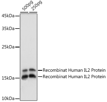
Western blot analysis of Recombinant Human IL2 Protein (RP01039), using IL2 Rabbit pAb (A16317) at 1:1000 dilution. Secondary antibody: HRP-conjugated Goat anti-Rabbit IgG (H+L) (AS014) at 1:10000 dilution. Lysates/proteins: 500pg per lane. Blocking buffer: 3% nonfat dry milk in TBST. Detection: ECL Basic Kit (RM00020). Exposure time: 90s. |
|
|
Western blot analysis of lysates from Recombinat Human IL2 Protein, using IL2 Rabbit pAb (A16317) at 1:1000 dilution. Secondary antibody: HRP-conjugated Goat anti-Rabbit IgG (H+L) (AS014) at 1:10000 dilution. Lysates/proteins: 25µg per lane. Blocking buffer: 3% nonfat dry milk in TBST. Detection: ECL Basic Kit (RM00020). Exposure time: 1s. |
|
|
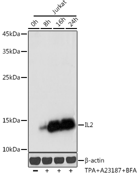
Western blot analysis of lysates from Jurkat cells, using IL2 Rabbit pAb (A16317) at 1:1000 dilution. Jurkat cells were treated with TPA (40 nM),A23187 (2µM) and Brefeldin A (300 ng / ml) for 0-24hours Secondary antibody: HRP-conjugated Goat anti-Rabbit IgG (H+L) (AS014) at 1:10000 dilution. Lysates/proteins: 25µg per lane. Blocking buffer: 3% nonfat dry milk in TBST. Detection: ECL Basic Kit (RM00020). Exposure time: 30s. |
|
|
Western blot analysis of lysates from Jurkat cells, using IL2 Rabbit pAb (A16898) at 1:1000 dilution. Jurkat cells were treated with TPA (40 nM),A23187 (2µM) and Brefeldin A (300 ng / ml) for 0-24hours Secondary antibody: HRP-conjugated Goat anti-Rabbit IgG (H+L) (AS014) at 1:10000 dilution. Lysates/proteins: 25µg per lane. Blocking buffer: 3% nonfat dry milk in TBST. Detection: ECL Basic Kit (RM00020). Exposure time: 1s. |
|
|
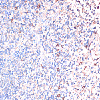
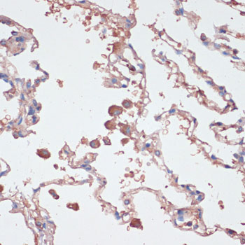
Immunohistochemistry analysis of paraffin-embedded Human tonsil using IL2 Rabbit pAb (A16317) at dilution of 1:100 (40x lens). Microwave antigen retrieval performed with 0.01M PBS Buffer (pH 7.2) prior to IHC staining. |
|
|
Immunohistochemistry analysis of paraffin-embedded Human lung using IL2 Rabbit pAb (A16317) at dilution of 1:100 (40x lens). Microwave antigen retrieval performed with 0.01M PBS Buffer (pH 7.2) prior to IHC staining. |
|
|
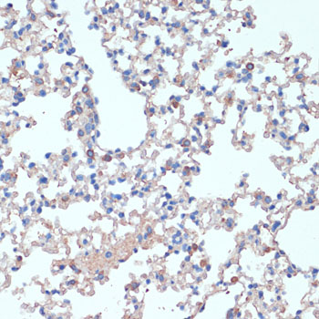
Immunohistochemistry analysis of paraffin-embedded Mouse lung using IL2 Rabbit pAb (A16317) at dilution of 1:100 (40x lens). Microwave antigen retrieval performed with 0.01M PBS Buffer (pH 7.2) prior to IHC staining. |
|
|
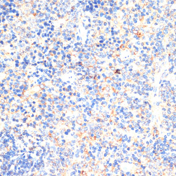
Immunohistochemistry analysis of paraffin-embedded Mouse spleen using IL2 Rabbit pAb (A16317) at dilution of 1:100 (40x lens). Microwave antigen retrieval performed with 0.01M PBS Buffer (pH 7.2) prior to IHC staining. |
Produktgarantie und fachkundiger Support
