
IFNAR2 Rabbit pAb, Unconjugated, Polyclonal
Artikelnummer:
ABB-A1769
- Bilder (8)

| Artikelname: | IFNAR2 Rabbit pAb, Unconjugated, Polyclonal |
| Artikelnummer: | ABB-A1769 |
| Hersteller Artikelnummer: | A1769 |
| Alternativnummer: | ABB-A1769-20UL,ABB-A1769-100UL,ABB-A1769-1000UL,ABB-A1769-500UL |
| Hersteller: | ABclonal |
| Wirt: | Rabbit |
| Kategorie: | Antikörper |
| Applikation: | ELISA, IF, IHC-P, WB |
| Spezies Reaktivität: | Human |
| Immunogen: | Recombinant protein (or fragment).This information is considered to be commercially sensitive. |
| Konjugation: | Unconjugated |
| Alternative Synonym: | IFN-R, IMD45, IFNABR, IFNARB, IFN-R-2, IFN-alpha-REC, IFNAR2 |
| The protein encoded by this gene is a type I membrane protein that forms one of the two chains of a receptor for interferons alpha and beta. Binding and activation of the receptor stimulates Janus protein kinases, which in turn phosphorylate several proteins, including STAT1 and STAT2. The protein belongs to the type II cytokine receptor family. Mutations in this gene are associated with Immunodeficiency 45. |
| Klonalität: | Polyclonal |
| Molekulargewicht: | 58kDa |
| NCBI: | 3455 |
| UniProt: | P48551 |
| Reinheit: | Affinity purification |
| Sequenz: | ISYDSPDYTDESCTFKISLRNFRSILSWELKNHSIVPTHYTLLYTIMSKPEDLKVVKNCANTTRSFCDLTDEWRSTHEAYVTVLEGFSGNTTLFSCSHNFWLAIDMSFEPPEFEIVGFTNHINVMVKFPSIVEEELQFDLSLVIEEQSEGIVKKHKPEIKGNMSGNFTYIIDKLIPNTNYCVSVYLEHSDEQAVIKSPLKCTLLPPGQESESAESAK |
| Target-Kategorie: | IFNAR2 |
| Antibody Type: | Primary Antibody |
| Application Verdünnung: | WB,1:1000 - 1:5000|IHC-P,1:50 - 1:100|IF/ICC,1:50 - 1:200|ELISA,Recommended starting concentration is 1 µg/mL. Please optimize the concentration based on your specific assay requirements. |
| Anwendungsbeschreibung: | Cross-Reactivity: Human,Mouse,Rat. ResearchArea: Epigenetics Nuclear Signaling,Cancer,Tumor immunology,Signal Transduction,Immunology Inflammation,Cytokines,Cell Intrinsic Innate Immunity Signaling Pathway. Shipping: Ice Bag |
|
|
Western blot analysis of various lysates using IFNAR2 Rabbit pAb (A1769) at 1:500 dilution incubated overnight at 4°C. Secondary antibody: HRP-conjugated Goat anti-Rabbit IgG (H+L) (AS014) at 1:10000 dilution. Lysates/proteins: 25 µg per lane. Blocking buffer: 3% nonfat dry milk in TBST. Detection: ECL Basic Kit (RM00020). Exposure time: 1s. |
|
|
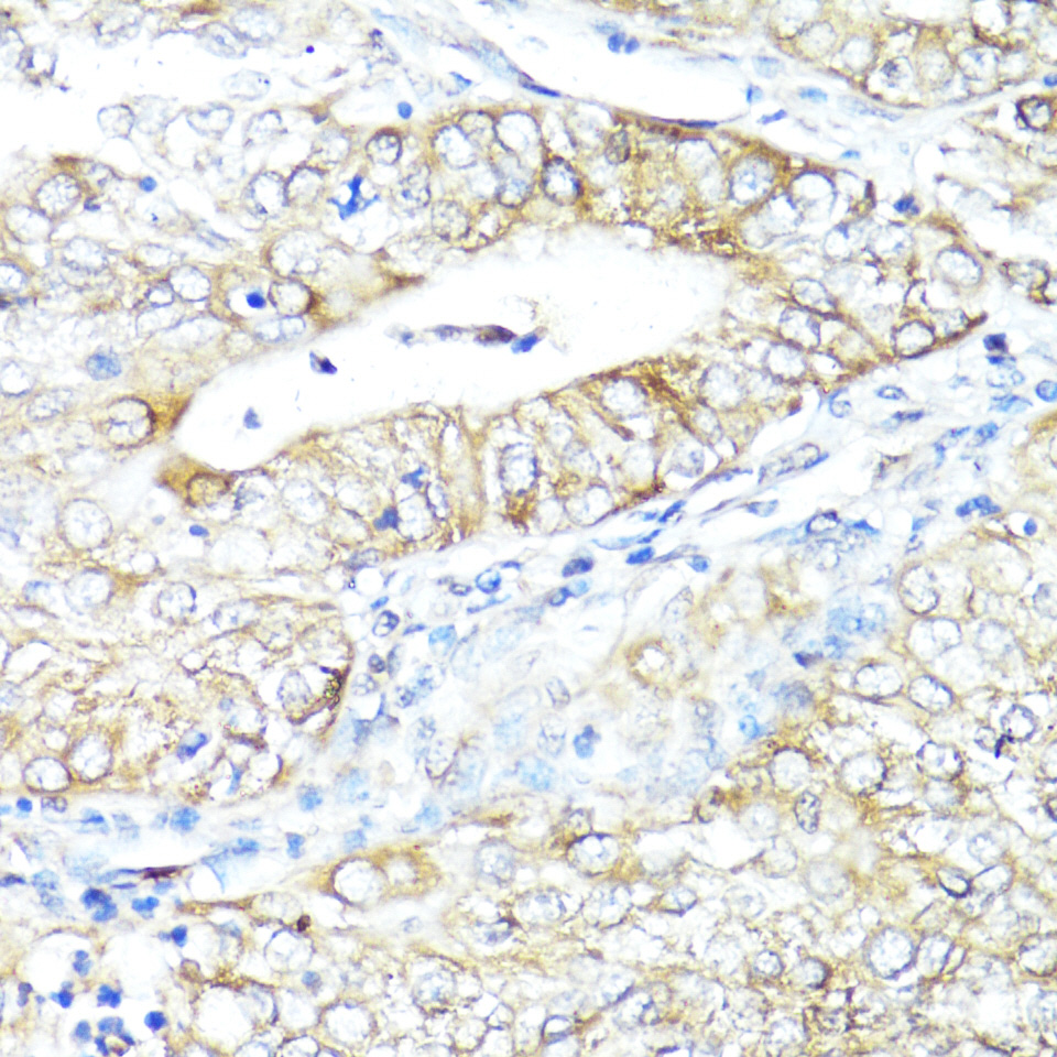
Immunohistochemistry analysis of paraffin-embedded Human uterine cancer using IFNAR2 Rabbit pAb (A1769) at dilution of 1:100 (40x lens). Microwave antigen retrieval performed with 0.01M PBS Buffer (pH 7.2) prior to IHC staining. |
|
|
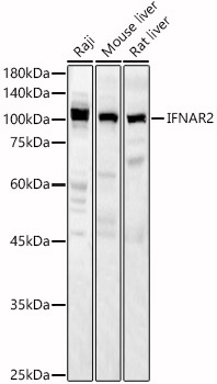
Western blot analysis of various lysates, using IFNAR2 Rabbit pAb (A1769) at 1:2000 dilution. Secondary antibody: HRP-conjugated Goat anti-Rabbit IgG (H+L) (AS014) at 1:10000 dilution. Lysates/proteins: 25µg per lane. Blocking buffer: 3% nonfat dry milk in TBST. Detection: ECL Basic Kit (RM00020). Exposure time: 60s. |
|
|
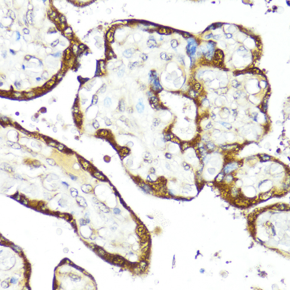
Immunohistochemistry analysis of paraffin-embedded Human placenta using IFNAR2 Rabbit pAb (A1769) at dilution of 1:100 (40x lens). Microwave antigen retrieval performed with 0.01M PBS Buffer (pH 7.2) prior to IHC staining. |
|
|
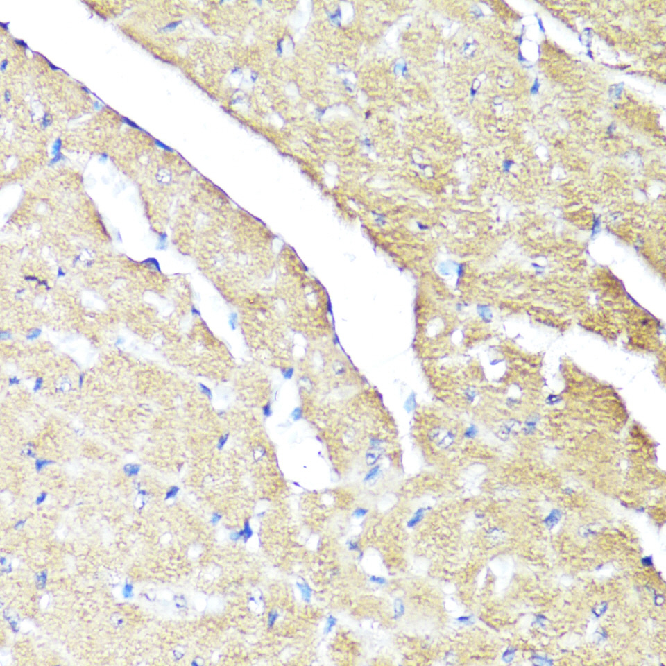
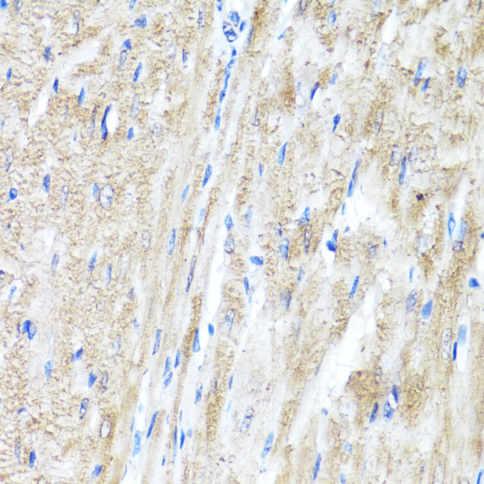
Immunohistochemistry analysis of paraffin-embedded Mouse heart using IFNAR2 Rabbit pAb (A1769) at dilution of 1:100 (40x lens). Microwave antigen retrieval performed with 0.01M PBS Buffer (pH 7.2) prior to IHC staining. |
|
|
Immunohistochemistry analysis of paraffin-embedded Rat heart using IFNAR2 Rabbit pAb (A1769) at dilution of 1:100 (40x lens). Microwave antigen retrieval performed with 0.01M PBS Buffer (pH 7.2) prior to IHC staining. |
|
|
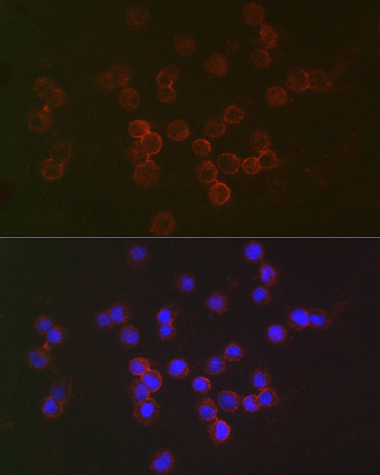
Immunofluorescence analysis of Raji cells using IFNAR2 Rabbit pAb (A1769) at dilution of 1:50 (40x lens). Secondary antibody: Cy3-conjugated Goat anti-Rabbit IgG (H+L) (AS007) at 1:500 dilution. Blue: DAPI for nuclear staining. |
|
|
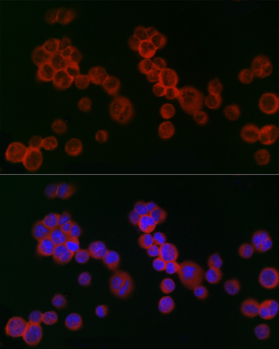
Immunofluorescence analysis of K-562 cells using IFNAR2 Rabbit pAb (A1769) at dilution of 1:100 (40x lens). Secondary antibody: Cy3-conjugated Goat anti-Rabbit IgG (H+L) (AS007) at 1:500 dilution. Blue: DAPI for nuclear staining. |
Produktgarantie und fachkundiger Support
