
[KO Validated] CD44 Rabbit mAb, Unconjugated, Monoclonal
Artikelnummer:
ABB-A19020
- Bilder (8)

| Artikelname: | [KO Validated] CD44 Rabbit mAb, Unconjugated, Monoclonal |
| Artikelnummer: | ABB-A19020 |
| Hersteller Artikelnummer: | A19020 |
| Alternativnummer: | ABB-A19020-100UL,ABB-A19020-20UL,ABB-A19020-1000UL,ABB-A19020-500UL |
| Hersteller: | ABclonal |
| Wirt: | Rabbit |
| Kategorie: | Antikörper |
| Applikation: | ELISA, FC, IF, IHC-P, WB |
| Spezies Reaktivität: | Human |
| Immunogen: | Recombinant protein (or fragment).This information is considered to be commercially sensitive. |
| Konjugation: | Unconjugated |
| Alternative Synonym: | IN, LHR, MC56, MDU2, MDU3, MIC4, Pgp1, CDW44, CSPG8, H-CAM, HCELL, ECM-III, HUTCH-1, HUTCH-I, ECMR-III, Hermes-1, CD44 |
| The protein encoded by this gene is a cell-surface glycoprotein involved in cell-cell interactions, cell adhesion and migration. It is a receptor for hyaluronic acid (HA) and can also interact with other ligands, such as osteopontin, collagens, and matrix metalloproteinases (MMPs). This protein participates in a wide variety of cellular functions including lymphocyte activation, recirculation and homing, hematopoiesis, and tumor metastasis. Transcripts for this gene undergo complex alternative splicing that results in many functionally distinct isoforms, however, the full length nature of some of these variants has not been determined. Alternative splicing is the basis for the structural and functional diversity of this protein, and may be related to tumor metastasis. |
| Klonalität: | Monoclonal |
| Klon-Bezeichnung: | [ARC52411] |
| Molekulargewicht: | 82kDa |
| NCBI: | 960 |
| UniProt: | P16070 |
| Reinheit: | Affinity purification |
| Sequenz: | AQIDLNITCRFAGVFHVEKNGRYSISRTEAADLCKAFNSTLPTMAQMEKALSIGFETCRYGFIEGHVVIPRIHPNSICAANNTGVYILTSNTSQYDTYCFNASAPPEEDCTSVTDLPNAFDGPITITIVNRDGTRYVQKGEYRTNPEDIYPSNPTDDDV |
| Target-Kategorie: | CD44 |
| Antibody Type: | Primary Antibody |
| Application Verdünnung: | WB,1:10000 - 1:40000|IHC-P,1:1000 - 1:5000|IF/ICC,1:200-1:2000|ELISA,Recommended starting concentration is 1 µg/mL. Please optimize the concentration based on your specific assay requirements. |
| Anwendungsbeschreibung: | Cross-Reactivity: Human. ResearchArea: Cancer,Tumor immunology,Tumor biomarkers,Immunology Inflammation,CDs,Stem Cells,Hematopoietic Progenitors,Mesenchymal Stem Cells. Shipping: Ice Bag |
|
|
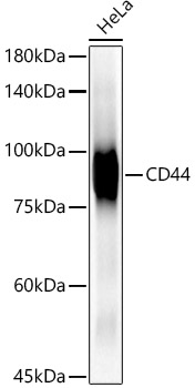
Western blot analysis of lysates from wild type (WT) and CD44 knockout (KO) HeLa cells using [KO Validated] CD44 Rabbit mAb at 1:24000 dilution incubated overnight at 4°C. Secondary antibody: HRP-conjugated Goat anti-Rabbit IgG (H+L) (AS014) at 1:10000 dilution. Lysates/proteins: 25 µg per lane. Blocking buffer: 3% nonfat dry milk in TBST. Detection: ECL Basic Kit (RM00020). Exposure time: 30s. |
|
|
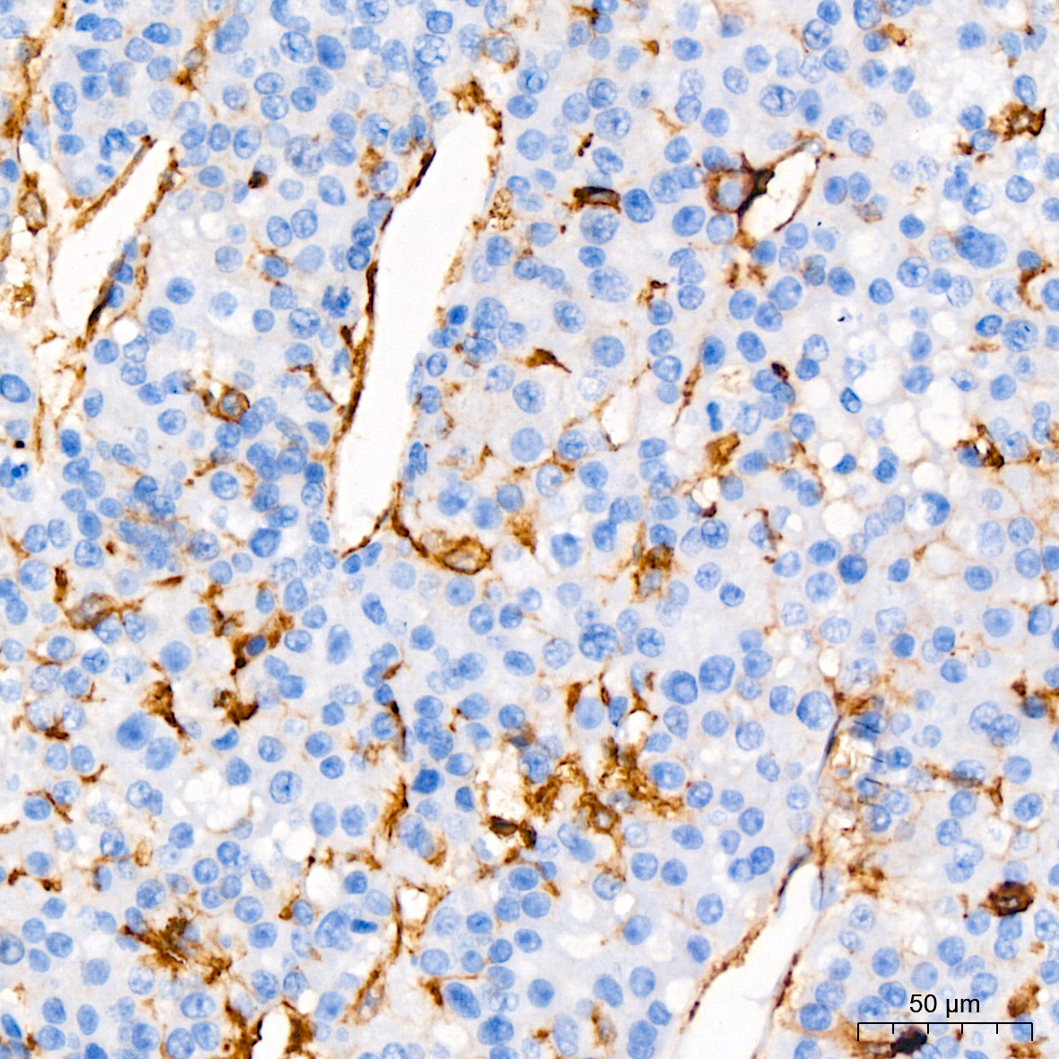
Immunohistochemistry analysis of paraffin-embedded Human liver cancer using [KO Validated] CD44 Rabbit mAb (A19020) at dilution of 1:2000 (40x lens). High pressure antigen retrieval performed with 0.01M Tris/EDTA Buffer (pH 9.0) prior to IHC staining. |
|
|
Western blot analysis of various lysates using [KO Validated] CD44 Rabbit mAb (A19020) at 1:20000 dilution incubated overnight at 4°C. Secondary antibody: HRP-conjugated Goat anti-Rabbit IgG (H+L) (AS014) at 1:10000 dilution. Lysates/proteins: 25 µg per lane. Blocking buffer: 3% nonfat dry milk in TBST. Detection: ECL Basic Kit (RM00020). Exposure time: 45s. |
|
|
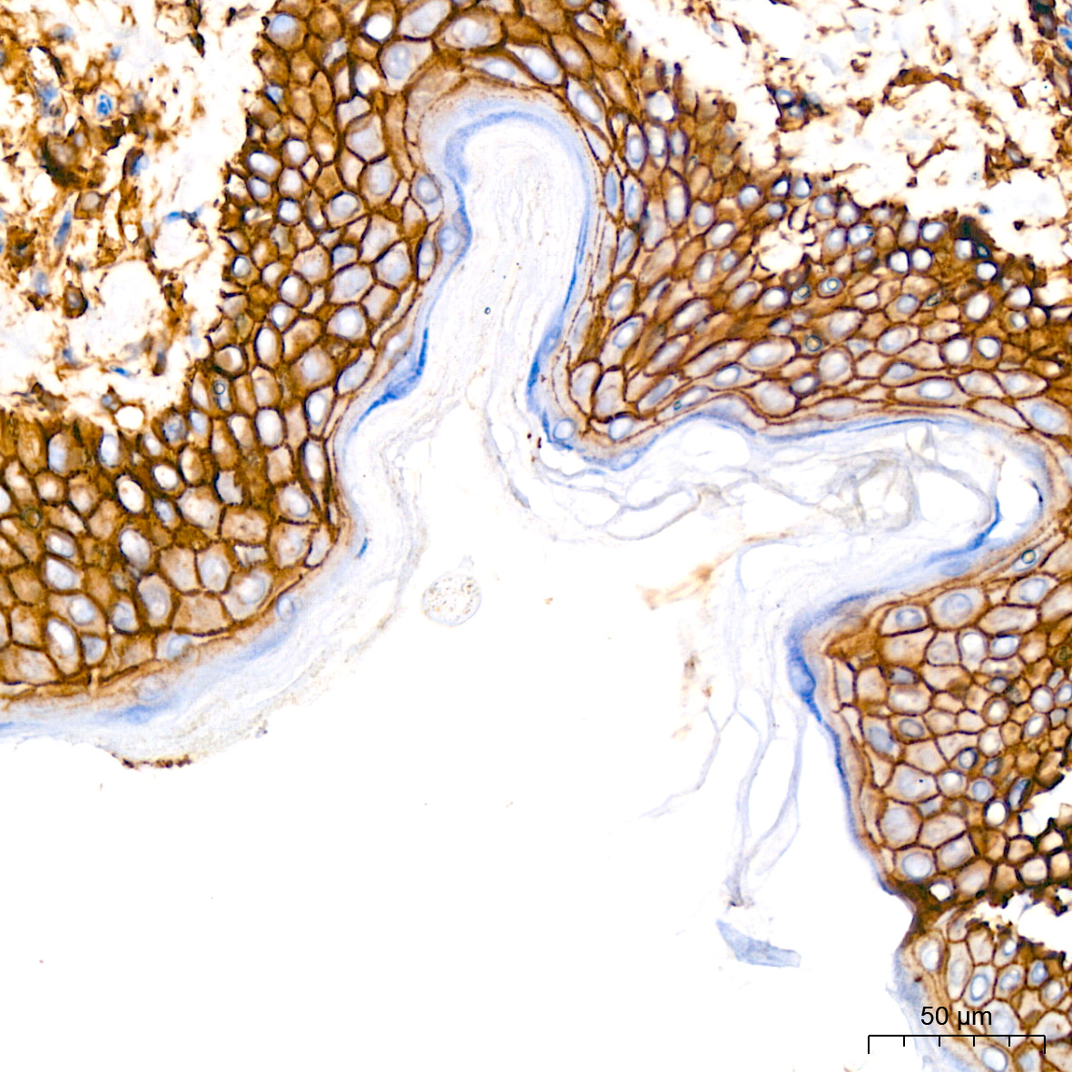
Immunohistochemistry analysis of paraffin-embedded Human skin using [KO Validated] CD44 Rabbit mAb (A19020) at dilution of 1:2000 (40x lens). High pressure antigen retrieval performed with 0.01M Tris/EDTA Buffer (pH 9.0) prior to IHC staining. |
|
|
Confocal imaging of A-431 cells using [KO Validated] CD44 Rabbit mAb (A19020, dilution 1:200) followed by a further incubation with Cy3 Goat Anti-Rabbit IgG (H+L) (AS007, dilution 1:500) (Red). The cells were counterstained with alpha-Tubulin Mouse mAb (AC012, dilution 1:400) followed by incubation with ABflo 488-conjugated Goat Anti-Mouse IgG (H+L) Ab (AS076, dilution 1:500) (Green). DAPI was used for nuclear staining (Blue). Objective: 100x. |
|
|
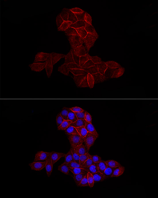
Confocal imaging of HeLa cells using [KO Validated] CD44 Rabbit mAb (A19020, dilution 1:200) followed by a further incubation with Cy3 Goat Anti-Rabbit IgG (H+L) (AS007, dilution 1:500) (Red). The cells were counterstained with alpha-Tubulin Mouse mAb (AC012, dilution 1:400) followed by incubation with ABflo 488-conjugated Goat Anti-Mouse IgG (H+L) Ab (AS076, dilution 1:500) (Green). DAPI was used for nuclear staining (Blue). Objective: 100x. |
|
|
Flow cytometry: 1X10 6 Human PBMC were surface-stained with Rabbit IgG isotype control (AC042,2 µg/mL,left) or [KO Validated] CD44 Rabbit mAb (A19020,2.5 µg/mL,right), followed by Alexa Fluor 647 conjugated goat anti-rabbit pAb staining. Cells in the lymphocyte gate were used for analysis. |
|
|
Flow cytometry: 1X10 6 HeLa cells were surface-stained with ABflo 647 Rabbit IgG isotype control (A22070,5 µl/Test,left) or [KO Validated] CD44 Rabbit mAb (A19020,2.5 µg/mL,right). |
Produktgarantie und fachkundiger Support
