
SHP1 Rabbit mAb, Unconjugated, Monoclonal
Artikelnummer:
ABB-A19111
- Bilder (8)

| Artikelname: | SHP1 Rabbit mAb, Unconjugated, Monoclonal |
| Artikelnummer: | ABB-A19111 |
| Hersteller Artikelnummer: | A19111 |
| Alternativnummer: | ABB-A19111-100UL,ABB-A19111-20UL |
| Hersteller: | ABclonal |
| Wirt: | Rabbit |
| Kategorie: | Antikörper |
| Applikation: | ELISA, IF, IHC-P, WB |
| Spezies Reaktivität: | Human |
| Immunogen: | Synthetic peptide. This information is considered to be commercially sensitive. |
| Konjugation: | Unconjugated |
| Alternative Synonym: | HCP, HCPH, SHP1, SHP-1, HPTP1C, PTP-1C, SHP-1L, SH-PTP1 |
| The protein encoded by this gene is a member of the protein tyrosine phosphatase (PTP) family. PTPs are known to be signaling molecules that regulate a variety of cellular processes including cell growth, differentiation, mitotic cycle, and oncogenic transformation. N-terminal part of this PTP contains two tandem Src homolog (SH2) domains, which act as protein phospho-tyrosine binding domains, and mediate the interaction of this PTP with its substrates. This PTP is expressed primarily in hematopoietic cells, and functions as an important regulator of multiple signaling pathways in hematopoietic cells. This PTP has been shown to interact with, and dephosphorylate a wide spectrum of phospho-proteins involved in hematopoietic cell signaling. Multiple alternatively spliced variants of this gene, which encode distinct isoforms, have been reported. |
| Application Verdünnung: | WB,1:1000 - 1:2000|IF-P,1:100 - 1:800|IHC-P,1:200 - 1:2000|ELISA,Recommended starting concentration is 1 µg/mL. Please optimize the concentration based on your specific assay requirements. |
| Anwendungsbeschreibung: | Cross-Reactivity: Human,Mouse,Rat. ResearchArea: Protein phosphorylation,Cancer,Signal Transduction,G protein signaling,G-Protein-Coupled ReceptorsGPCR,Kinase,Tyrosine kinases,Cell Biology Developmental Biology,Apoptosis,Cell Cycle,Immunology Inflammation,B Cell Receptor Signaling Pathway,Jak-Stat-IL-6 Receptor Signaling Pathway. Shipping: Ice Bag |
|
|
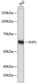
Western blot analysis of lysates from Raji cells, using SHP1 Rabbit mAb (A19111) at 1:1000 dilution. Secondary antibody: HRP-conjugated Goat anti-Rabbit IgG (H+L) (AS014) at 1:10000 dilution. Lysates/proteins: 25µg per lane. Blocking buffer: 3% nonfat dry milk in TBST. Detection: ECL Basic Kit (RM00020). Exposure time: 10s. |
|
|
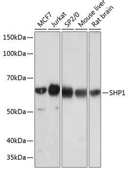
Western blot analysis of various lysates using SHP1 Rabbit mAb (A19111) at 1:1000 dilution. Secondary antibody: HRP-conjugated Goat anti-Rabbit IgG (H+L) (AS014) at 1:10000 dilution. Lysates/proteins: 25µg per lane. Blocking buffer: 3% nonfat dry milk in TBST. Detection: ECL Basic Kit (RM00020). Exposure time: 3min. |
|
|
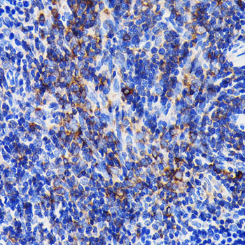
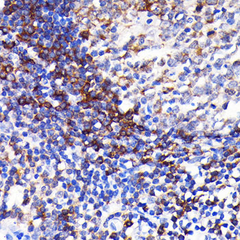
Immunohistochemistry analysis of paraffin-embedded Human spleen tissue using SHP1 Rabbit mAb (A19111) at a dilution of 1:200 (40x lens). High pressure antigen retrieval performed with 0.01M Citrate buffer (pH 6.0) prior to IHC staining. |
|
|
Immunohistochemistry analysis of paraffin-embedded Mouse spleen tissue using SHP1 Rabbit mAb (A19111) at a dilution of 1:800 (40x lens). High pressure antigen retrieval performed with 0.01M Citrate buffer (pH 6.0) prior to IHC staining. |
|
|
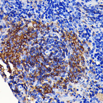
Immunohistochemistry analysis of paraffin-embedded Human tonsil tissue using SHP1 Rabbit mAb (A19111) at a dilution of 1:200 (40x lens). High pressure antigen retrieval performed with 0.01M Citrate buffer (pH 6.0) prior to IHC staining. |
|
|
Immunohistochemistry analysis of paraffin-embedded Rat spleen tissue using SHP1 Rabbit mAb (A19111) at a dilution of 1:800 (40x lens). High pressure antigen retrieval performed with 0.01M Citrate buffer (pH 6.0) prior to IHC staining. |
|
|
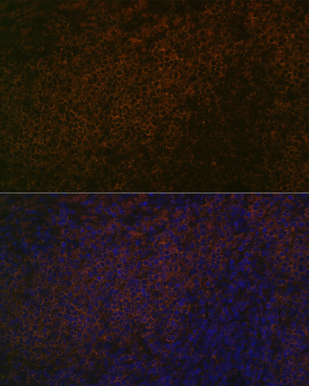
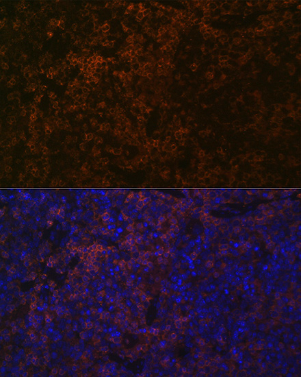
Immunofluorescence analysis of paraffin-embedded rat spleen using SHP1 Rabbit mAb (A19111) at dilution of 1:100 (40x lens). Secondary antibody: Cy3-conjugated Goat anti-Rabbit IgG (H+L) (AS007) at 1:500 dilution. Blue: DAPI for nuclear staining. |
|
|
Immunofluorescence analysis of paraffin-embedded mouse spleen using SHP1 Rabbit mAb (A19111) at dilution of 1:100 (40x lens). Secondary antibody: Cy3-conjugated Goat anti-Rabbit IgG (H+L) (AS007) at 1:500 dilution. Blue: DAPI for nuclear staining. |
Produktgarantie und fachkundiger Support
