
[KD Validated] ERK1 Rabbit mAb, Unconjugated, Monoclonal
Artikelnummer:
ABB-A19561
- Bilder (8)

| Artikelname: | [KD Validated] ERK1 Rabbit mAb, Unconjugated, Monoclonal |
| Artikelnummer: | ABB-A19561 |
| Hersteller Artikelnummer: | A19561 |
| Alternativnummer: | ABB-A19561-20UL,ABB-A19561-100UL,ABB-A19561-1000UL,ABB-A19561-500UL |
| Hersteller: | ABclonal |
| Wirt: | Rabbit |
| Kategorie: | Antikörper |
| Applikation: | ELISA, IF, IHC-P, IP, WB |
| Spezies Reaktivität: | Human |
| Immunogen: | Synthetic peptide. This information is considered to be commercially sensitive. |
| Konjugation: | Unconjugated |
| Alternative Synonym: | ERK1, ERT2, ERK-1, PRKM3, P44ERK1, P44MAPK, HS44KDAP, HUMKER1A, p44-ERK1, p44-MAPK, K1 |
| The protein encoded by this gene is a member of the MAP kinase family. MAP kinases, also known as extracellular signal-regulated kinases (ERKs), act in a signaling cascade that regulates various cellular processes such as proliferation, differentiation, and cell cycle progression in response to a variety of extracellular signals. This kinase is activated by upstream kinases, resulting in its translocation to the nucleus where it phosphorylates nuclear targets. Alternatively spliced transcript variants encoding different protein isoforms have been described. |
| Application Verdünnung: | WB,1:1000 - 1:6000|IP,0.5µg-4µg antibody for 200µg-400µg extracts of whole cells|IF/ICC,1:100 - 1:800|IF-P,1:100 - 1:800|IHC-P,1:200 - 1:2000|ELISA,Recommended starting concentration is 1 µg/mL. Please optimize the concentration based on your specific ass |
| Anwendungsbeschreibung: | Cross-Reactivity: Human,Mouse. ResearchArea: Epigenetics Nuclear Signaling,Translation Control,Regulation of eIF4 and p70 S6 Kinase,Protein phosphorylation,Signal Transduction,G protein signaling,G-Protein-Coupled Receptors Signaling to MAPK Erk,Kinase,Serine threonine kinases,mTOR Signaling Pathway,ErbB-HER Signaling Pathway,MAPK-Erk Signaling Pathway,Cell Biology Developmental Biology,Apoptosis,Mitochondrial Control of Apoptosis,Inhibition of Apoptosis,Cell Cycle,Microtubules,TGF-b-Smad Signaling Pathway,ESC Pluripotency and Differentiation,Endocrine Metabolism,Insulin Receptor Signaling Pathway,Warburg Effect,Immunology Inflammation,B Cell Receptor Signaling Pathway,T Cell Receptor Signaling Pathway,Jak-Stat-IL-6 Receptor Signaling Pathway,Neuroscience,Neurodegenerative Diseases,Amyloid Plaque and Neurofibrillary Tangle Formation in Alzheimers Disease,Stem Cells,Cardiovascular,Angiogenesis. Shipping: Ice Bag |
|
|
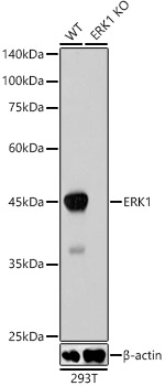
Western blot analysis of lysates from wild type(WT) and ERK1 knockdown (KD) 293T cells, using [KD Validated] ERK1 Rabbit mAb (A19561) at 1:1000 dilution. Secondary antibody: HRP-conjugated Goat anti-Rabbit IgG (H+L) (AS014) at 1:10000 dilution. Lysates/proteins: 25µg per lane. Blocking buffer: 3% nonfat dry milk in TBST. Detection: ECL Basic Kit (RM00020). Exposure time: 1s. |
|
|
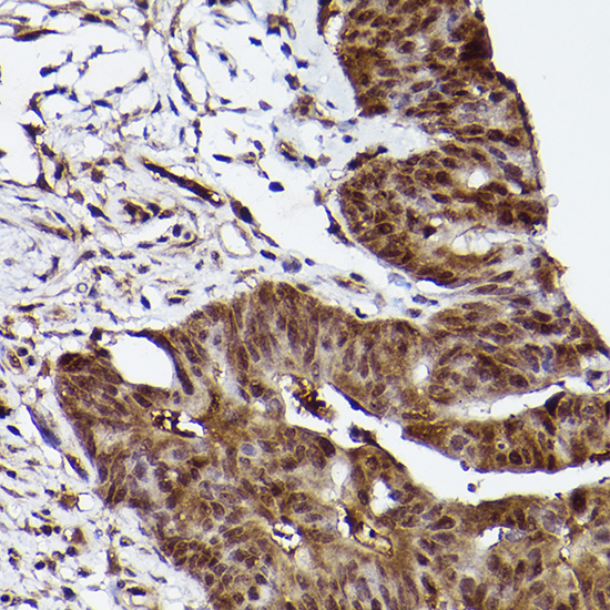
Immunohistochemistry analysis of paraffin-embeddedHuman thyroid cancer tissue using[KD Validated] ERK1 Rabbit mAb(A19561) at a dilution of 1:200 (40x lens).High pressure antigen retrieval was performed with 0.01 M citrate buffer (pH 6.0) prior to IHC staining. |
|
|
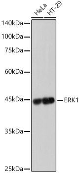
Western blot analysis of various lysates, using [KD Validated] ERK1 Rabbit mAb (A19561) at 1:1000 dilution. Secondary antibody: HRP-conjugated Goat anti-Rabbit IgG (H+L) (AS014) at 1:10000 dilution. Lysates/proteins: 25µg per lane. Blocking buffer: 3% nonfat dry milk in TBST. Detection: ECL Basic Kit (RM00020). Exposure time: 1s. |
|
|
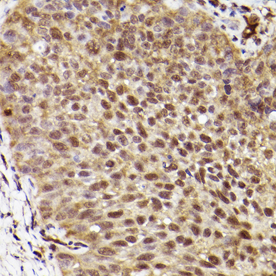
Immunohistochemistry analysis of paraffin-embeddedHuman brain tissue using[KD Validated] ERK1 Rabbit mAb(A19561) at a dilution of 1:200 (40x lens).High pressure antigen retrieval was performed with 0.01 M citrate buffer (pH 6.0) prior to IHC staining. |
|
|
Immunohistochemistry analysis of paraffin-embeddedHuman breast tissue using[KD Validated] ERK1 Rabbit mAb(A19561) at a dilution of 1:200 (40x lens).High pressure antigen retrieval was performed with 0.01 M citrate buffer (pH 6.0) prior to IHC staining. |
|
|
Confocal imaging of MCF7 cells using[KD Validated] ERK1 Rabbit mAb (A19561, dilution 1:100) followed by a further incubation with Cy3 Goat Anti-Rabbit IgG (H+L) (AS007, dilution 1:500) (Red). The cells were counterstained with alpha-Tubulin Mouse mAb (AC012, dilution 1:400) followed by incubation with ABflo 488-conjugated Goat Anti-Mouse IgG (H+L) Ab (AS076, dilution 1:500) (Green). DAPI was used for nuclear staining (Blue). Objective: 100x. |
|
|
Confocal imaging ofparaffin-embedded 5xFAD mouse brain tissue using[KD Validated] ERK1 Rabbit mAb (A19561, dilution 1:100) followed by a further incubation with Cy3 Goat Anti-Rabbit IgG (H+L) (AS007, dilution 1:500) (Red). DAPI was used for nuclear staining (Blue). Objective: 40x. Perform microwave antigen retrieval with 0.01 M citrate buffer (pH 6.0) prior to IF staining. |
|
|
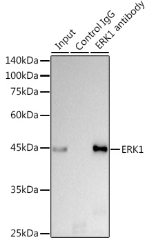
Immunoprecipitation analysis of 300 µg extracts of HeLa cells using 3 µg [KD Validated] ERK1 Rabbit mAb (A19561). Western blot was performed from the immunoprecipitate using ERK1 antibody (A19561) at a dilution of 1:1000. |
Produktgarantie und fachkundiger Support
