
TACC3 Rabbit mAb, Unconjugated, Monoclonal
Artikelnummer:
ABB-A19617
- Bilder (8)


| Artikelname: | TACC3 Rabbit mAb, Unconjugated, Monoclonal |
| Artikelnummer: | ABB-A19617 |
| Hersteller Artikelnummer: | A19617 |
| Alternativnummer: | ABB-A19617-20UL,ABB-A19617-100UL |
| Hersteller: | ABclonal |
| Wirt: | Rabbit |
| Kategorie: | Antikörper |
| Applikation: | ELISA, IF, IHC-P, WB |
| Spezies Reaktivität: | Human |
| Immunogen: | Synthetic peptide. This information is considered to be commercially sensitive. |
| Konjugation: | Unconjugated |
| Alternative Synonym: | ERIC1, Tacc4, ERIC-1, maskin, TACC3 |
| TACC3, also known as ERIC 1, belongs to the TACC family. TACC family members TACC1, TACC2, and TACC3 map very closely to the corresponding FGFR1, FGFR2, FGFR3 genes on chromosomes 8, 10, and 4. Subsequently, since they are phylogenetically related, it is proposed that TACC and FGFR have similar roles in cell growth and differentiation. It plays a role in the microtubule-dependent coupling of the nucleus and the centrosome and involved in the processes that regulate centrosome-mediated interkinetic nuclear migration (INM) of neural progenitors. |
| Application Verdünnung: | WB,1:500 - 1:1000|IHC-P,1:50 - 1:200|IF/ICC,1:50 - 1:200|ELISA,Recommended starting concentration is 1 µg/mL. Please optimize the concentration based on your specific assay requirements. |
| Anwendungsbeschreibung: | Cross-Reactivity: Human,Mouse,Rat. ResearchArea: Cell Biology Developmental Biology,Apoptosis,Cell Cycle,Cell differentiation. Shipping: Ice Bag |
|
|
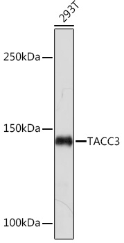
Western blot analysis of lysates from 293T cells, using TACC3 Rabbit mAb (A19617) at 1:1000 dilution. Secondary antibody: HRP-conjugated Goat anti-Rabbit IgG (H+L) (AS014) at 1:10000 dilution. Lysates/proteins: 25µg per lane. Blocking buffer: 3% nonfat dry milk in TBST. Detection: ECL Basic Kit (RM00020). Exposure time: 30s. |
|
|
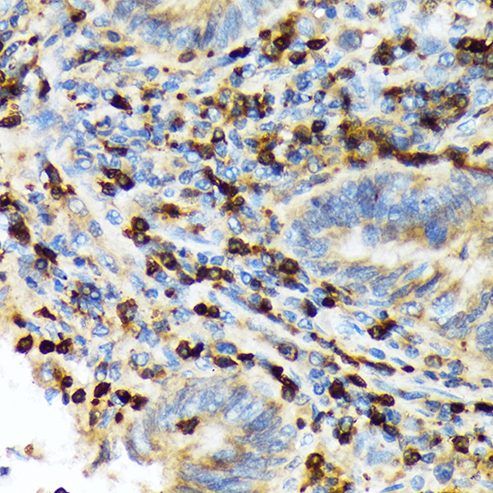
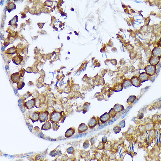
Immunohistochemistry analysis of paraffin-embeddedHuman colon tissue usingTACC3 Rabbit mAb(A19617) at a dilution of 1:200 (40x lens).High pressure antigen retrieval was performed with 0.01 M citrate buffer (pH 6.0) prior to IHC staining. |
|
|
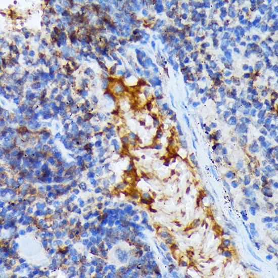
Immunohistochemistry analysis of paraffin-embeddedHuman tonsil tissue usingTACC3 Rabbit mAb(A19617) at a dilution of 1:200 (40x lens).High pressure antigen retrieval was performed with 0.01 M citrate buffer (pH 6.0) prior to IHC staining. |
|
|
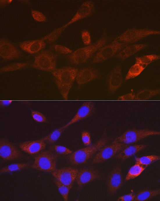
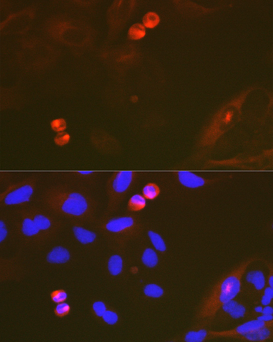
Immunofluorescence analysis of U-2 OS cells using TACC3 Rabbit mAb (A19617) at a dilution of 1:100 (40x lens). Secondary antibody: Cy3-conjugated Goat anti-Rabbit IgG (H+L)(AS007) at 1:500 dilution. Blue: DAPI for nuclear staining. |
|
|
Immunofluorescence analysis of NIH/3T3 cells using TACC3 Rabbit mAb (A19617) at a dilution of 1:100 (40x lens). Secondary antibody: Cy3-conjugated Goat anti-Rabbit IgG (H+L)(AS007) at 1:500 dilution. Blue: DAPI for nuclear staining. |
|
|
Immunofluorescence analysis of C6 cells using TACC3 Rabbit mAb (A19617) at a dilution of 1:100 (40x lens). Secondary antibody: Cy3-conjugated Goat anti-Rabbit IgG (H+L)(AS007) at 1:500 dilution. Blue: DAPI for nuclear staining. |
|
|
Immunofluorescence analysis of NIH/3T3 cells using TACC3 Rabbit mAb (A19617) at a dilution of 1:100 (40x lens). Secondary antibody: Cy3-conjugated Goat anti-Rabbit IgG (H+L)(AS007) at 1:500 dilution. Blue: DAPI for nuclear staining. |
|
|
Immunofluorescence analysis of C6 cells using TACC3 Rabbit mAb (A19617) at a dilution of 1:100 (40x lens). Secondary antibody: Cy3-conjugated Goat anti-Rabbit IgG (H+L)(AS007) at 1:500 dilution. Blue: DAPI for nuclear staining. |
Produktgarantie und fachkundiger Support
