
COPA Rabbit mAb, Unconjugated, Monoclonal
Artikelnummer:
ABB-A19651
- Bilder (8)

| Artikelname: | COPA Rabbit mAb, Unconjugated, Monoclonal |
| Artikelnummer: | ABB-A19651 |
| Hersteller Artikelnummer: | A19651 |
| Alternativnummer: | ABB-A19651-100UL,ABB-A19651-20UL |
| Hersteller: | ABclonal |
| Wirt: | Rabbit |
| Kategorie: | Antikörper |
| Applikation: | ELISA, IHC-P, WB |
| Spezies Reaktivität: | Human |
| Immunogen: | Synthetic peptide. This information is considered to be commercially sensitive. |
| Konjugation: | Unconjugated |
| Alternative Synonym: | AILJK, HEP-COP, alpha-COP, COPA |
| In eukaryotic cells, protein transport between the endoplasmic reticulum and Golgi compartments is mediated in part by non-clathrin-coated vesicular coat proteins (COPs). Seven coat proteins have been identified, and they represent subunits of a complex known as coatomer. The subunits are designated alpha-COP, beta-COP, beta-prime-COP, gamma-COP, delta-COP, epsilon-COP, and zeta-COP. The alpha-COP, encoded by COPA, shares high sequence similarity with RET1P, the alpha subunit of the coatomer complex in yeast. Also, the N-terminal 25 amino acids of alpha-COP encode the bioactive peptide, xenin, which stimulates exocrine pancreatic secretion and may act as a gastrointestinal hormone. Alternative splicing results in multiple splice forms encoding distinct isoforms. |
| Application Verdünnung: | WB,1:500 - 1:1000|IHC-P,1:50 - 1:200|ELISA,Recommended starting concentration is 1 µg/mL. Please optimize the concentration based on your specific assay requirements. |
| Anwendungsbeschreibung: | Cross-Reactivity: Human,Mouse,Rat. ResearchArea: Signal Transduction. Shipping: Ice Bag |
|
|
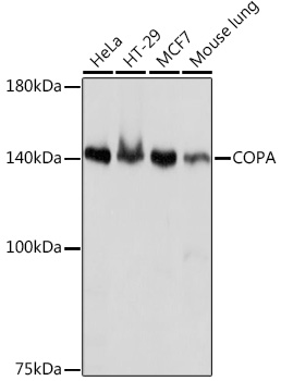
Western blot analysis of various lysates using COPA Rabbit mAb (A19651) at 1:1000 dilution. Secondary antibody: HRP-conjugated Goat anti-Rabbit IgG (H+L) (AS014) at 1:10000 dilution. Lysates/proteins: 25µg per lane. Blocking buffer: 3% nonfat dry milk in TBST. Detection: ECL Basic Kit (RM00020). Exposure time: 1s. |
|
|
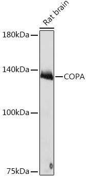
Western blot analysis of lysates from Rat brain, using COPA Rabbit mAb (A19651) at 1:1000 dilution. Secondary antibody: HRP-conjugated Goat anti-Rabbit IgG (H+L) (AS014) at 1:10000 dilution. Lysates/proteins: 25µg per lane. Blocking buffer: 3% nonfat dry milk in TBST. Detection: ECL Basic Kit (RM00020). Exposure time: 10s. |
|
|
Immunohistochemistry analysis of paraffin-embeddedRat colon tissue usingCOPA Rabbit mAb(A19651) at a dilution of 1:200 (40x lens).High pressure antigen retrieval was performed with 0.01 M citrate buffer (pH 6.0) prior to IHC staining. |
|
|
Immunohistochemistry analysis of paraffin-embeddedMouse testis tissue usingCOPA Rabbit mAb(A19651) at a dilution of 1:200 (40x lens).High pressure antigen retrieval was performed with 0.01 M citrate buffer (pH 6.0) prior to IHC staining. |
|
|
Immunohistochemistry analysis of paraffin-embeddedHuman thyroid cancer tissue usingCOPA Rabbit mAb(A19651) at a dilution of 1:200 (40x lens).High pressure antigen retrieval was performed with 0.01 M citrate buffer (pH 6.0) prior to IHC staining. |
|
|
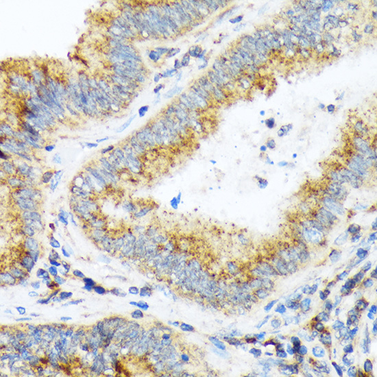
Immunohistochemistry analysis of paraffin-embeddedHuman colon tissue usingCOPA Rabbit mAb(A19651) at a dilution of 1:200 (40x lens).High pressure antigen retrieval was performed with 0.01 M citrate buffer (pH 6.0) prior to IHC staining. |
|
|
Immunohistochemistry analysis of paraffin-embeddedHuman colon carcinoma tissue usingCOPA Rabbit mAb(A19651) at a dilution of 1:200 (40x lens).High pressure antigen retrieval was performed with 0.01 M citrate buffer (pH 6.0) prior to IHC staining. |
|
|
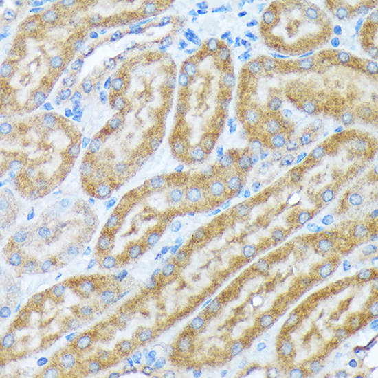
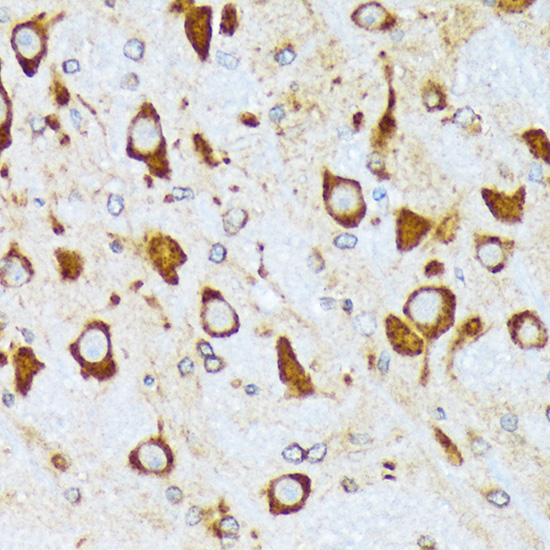
Immunohistochemistry analysis of paraffin-embeddedMouse brain tissue usingCOPA Rabbit mAb(A19651) at a dilution of 1:200 (40x lens).High pressure antigen retrieval was performed with 0.01 M citrate buffer (pH 6.0) prior to IHC staining. |
Produktgarantie und fachkundiger Support
