
NDUFS1 Rabbit mAb, Unconjugated, Monoclonal
Artikelnummer:
ABB-A21192
- Bilder (8)

| Artikelname: | NDUFS1 Rabbit mAb, Unconjugated, Monoclonal |
| Artikelnummer: | ABB-A21192 |
| Hersteller Artikelnummer: | A21192 |
| Alternativnummer: | ABB-A21192-20UL,ABB-A21192-100UL,ABB-A21192-1000UL,ABB-A21192-500UL |
| Hersteller: | ABclonal |
| Wirt: | Rabbit |
| Kategorie: | Antikörper |
| Applikation: | ELISA, IHC-P, WB |
| Spezies Reaktivität: | Human |
| Immunogen: | Recombinant protein (or fragment).This information is considered to be commercially sensitive. |
| Konjugation: | Unconjugated |
| Alternative Synonym: | CI-75k, MC1DN5, CI-75Kd, PRO1304, NDUFS1 |
| The protein encoded by this gene belongs to the complex I 75 kDa subunit family. Mammalian complex I is composed of 45 different subunits. It locates at the mitochondrial inner membrane. This protein has NADH dehydrogenase activity and oxidoreductase activity. It transfers electrons from NADH to the respiratory chain. The immediate electron acceptor for the enzyme is believed to be ubiquinone. This protein is the largest subunit of complex I and it is a component of the iron-sulfur (IP) fragment of the enzyme. It may form part of the active site crevice where NADH is oxidized. Mutations in this gene are associated with complex I deficiency. Several transcript variants encoding different isoforms have been found for this gene. |
| Klonalität: | Monoclonal |
| Klon-Bezeichnung: | [ARC52613] |
| Molekulargewicht: | 79kDa |
| NCBI: | 4719 |
| UniProt: | P28331 |
| Reinheit: | Affinity purification |
| Sequenz: | VEIEKAPKVVAACAMPVMKGWNILTNSEKSKKAREGVMEFLLANHPLDCPICDQGGECDLQDQSMMFGNDRSRFLEGKRAVEDKNIGPLVKTIMTRCIQCTRCIRFASEIAGVDDLGTTGRGNDMQVGTYIEKMFMSELSGNIIDICPVGALTSKPYAFTARPWETRKTESIDVMDAVGSNIVVSTRTGEVMRILPRMHEDINEEWISDKT |
| Target-Kategorie: | NDUFS1 |
| Antibody Type: | Primary Antibody |
| Application Verdünnung: | WB,1:1000 - 1:5000|IHC-P,1:100 - 1:500|ELISA,Recommended starting concentration is 1 µg/mL. Please optimize the concentration based on your specific assay requirements. |
| Anwendungsbeschreibung: | Cross-Reactivity: Human,Mouse,Rat. ResearchArea: Cancer,Cell Biology Developmental Biology,Apoptosis,Endocrine Metabolism,Mitochondrial metabolism,Mitochondrial markers,Oxidative phosphorylation,Neuroscience,Neurodegenerative Diseases. Shipping: Ice Bag |
|
|
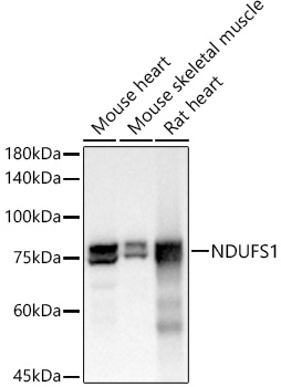
Western blot analysis of various lysates using NDUFS1 Rabbit mAb (A21192) at 1:2000 dilution. Secondary antibody: HRP-conjugated Goat anti-Rabbit IgG (H+L) (AS014) at 1:10000 dilution. Lysates/proteins: 25µg per lane. Blocking buffer: 3% nonfat dry milk in TBST. Detection: ECL Basic Kit (RM00020). Exposure time: 0.5s. |
|
|
Immunohistochemistry analysis of paraffin-embedded Human colon tissue using NDUFS1 Rabbit mAb (A21192) at a dilution of 1:400 (40x lens). High pressure antigen retrieval performed with 0.01M Citrate buffer (pH 6.0) prior to IHC staining. |
|
|
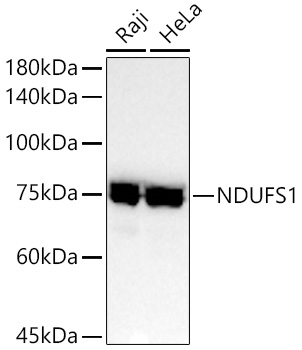
Western blot analysis of various lysates using NDUFS1 Rabbit mAb (A21192) at 1:2000 dilution. Secondary antibody: HRP-conjugated Goat anti-Rabbit IgG (H+L) (AS014) at 1:10000 dilution. Lysates/proteins: 25µg per lane. Blocking buffer: 3% nonfat dry milk in TBST. Detection: ECL Basic Kit (RM00020). Exposure time: 10s. |
|
|
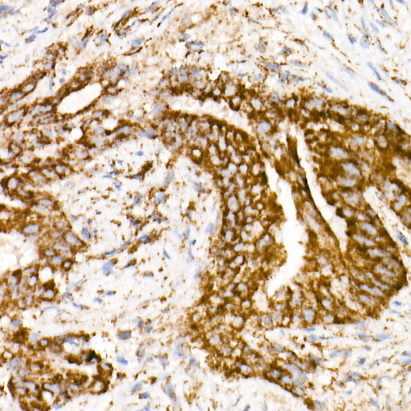
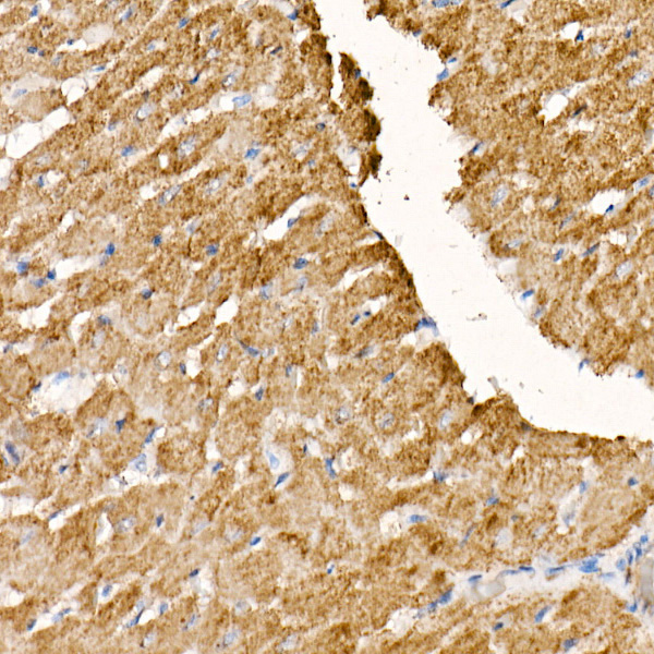
Immunohistochemistry analysis of paraffin-embedded Human liver cancer tissue using NDUFS1 Rabbit mAb (A21192) at a dilution of 1:400 (40x lens). High pressure antigen retrieval performed with 0.01M Citrate buffer (pH 6.0) prior to IHC staining. |
|
|
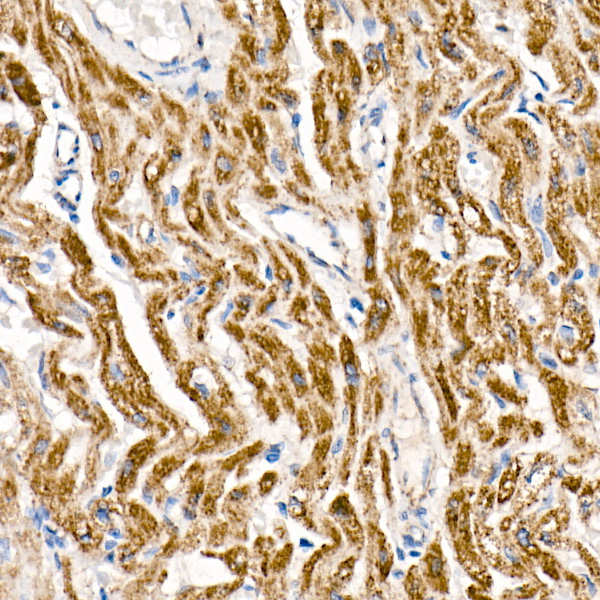
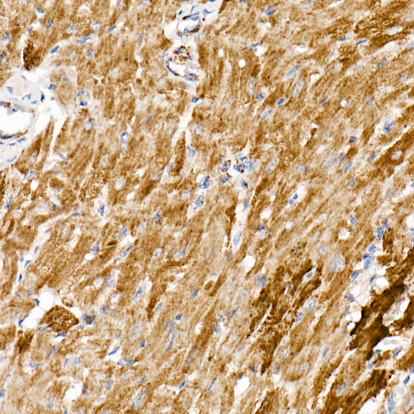
Immunohistochemistry analysis of paraffin-embedded Human liver tissue using NDUFS1 Rabbit mAb (A21192) at a dilution of 1:400 (40x lens). High pressure antigen retrieval performed with 0.01M Citrate buffer (pH 6.0) prior to IHC staining. |
|
|
Immunohistochemistry analysis of paraffin-embedded Human lung adenocarcinoma tissue using NDUFS1 Rabbit mAb (A21192) at a dilution of 1:400 (40x lens). High pressure antigen retrieval performed with 0.01M Citrate buffer (pH 6.0) prior to IHC staining. |
|
|
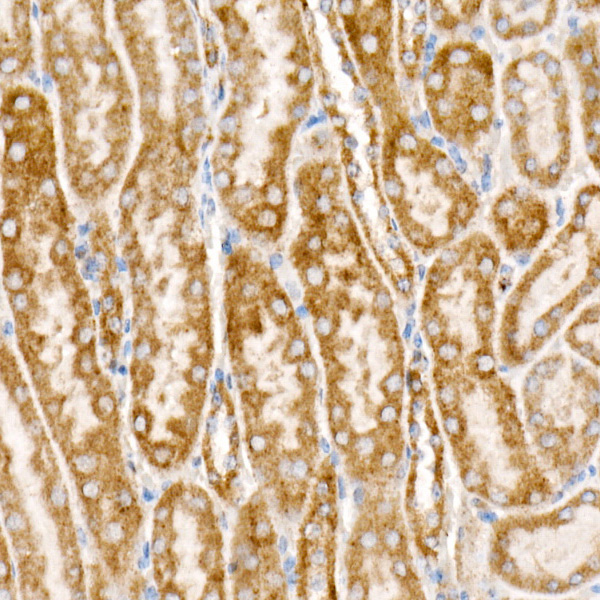
Immunohistochemistry analysis of paraffin-embedded Mouse kidney tissue using NDUFS1 Rabbit mAb (A21192) at a dilution of 1:400 (40x lens). High pressure antigen retrieval performed with 0.01M Citrate buffer (pH 6.0) prior to IHC staining. |
|
|
Immunohistochemistry analysis of paraffin-embedded Rat kidney tissue using NDUFS1 Rabbit mAb (A21192) at a dilution of 1:400 (40x lens). High pressure antigen retrieval performed with 0.01M Citrate buffer (pH 6.0) prior to IHC staining. |
Produktgarantie und fachkundiger Support
