
TNFSF10 Rabbit pAb, Unconjugated, Polyclonal
Artikelnummer:
ABB-A2138
- Bilder (1)
| Artikelname: | TNFSF10 Rabbit pAb, Unconjugated, Polyclonal |
| Artikelnummer: | ABB-A2138 |
| Hersteller Artikelnummer: | A2138 |
| Alternativnummer: | ABB-A2138-100UL,ABB-A2138-20UL,ABB-A2138-1000UL,ABB-A2138-500UL |
| Hersteller: | ABclonal |
| Wirt: | Rabbit |
| Kategorie: | Antikörper |
| Applikation: | ELISA, WB |
| Spezies Reaktivität: | Human |
| Immunogen: | Recombinant protein (or fragment).This information is considered to be commercially sensitive. |
| Konjugation: | Unconjugated |
| Alternative Synonym: | TL2, APO2L, CD253, TANCR, TRAIL, Apo-2L, TNLG6A, TNFSF10 |
| The protein encoded by this gene is a cytokine that belongs to the tumor necrosis factor (TNF) ligand family. This protein preferentially induces apoptosis in transformed and tumor cells, but does not appear to kill normal cells although it is expressed at a significant level in most normal tissues. This protein binds to several members of TNF receptor superfamily including TNFRSF10A/TRAILR1, TNFRSF10B/TRAILR2, TNFRSF10C/TRAILR3, TNFRSF10D/TRAILR4, and possibly also to TNFRSF11B/OPG. The activity of this protein may be modulated by binding to the decoy receptors TNFRSF10C/TRAILR3, TNFRSF10D/TRAILR4, and TNFRSF11B/OPG that cannot induce apoptosis. The binding of this protein to its receptors has been shown to trigger the activation of MAPK8/JNK, caspase 8, and caspase 3. Alternatively spliced transcript variants encoding different isoforms have been found for this gene. |
| Application Verdünnung: | WB,1:500 - 1:1000|ELISA,Recommended starting concentration is 1 µg/mL. Please optimize the concentration based on your specific assay requirements. |
| Anwendungsbeschreibung: | Cross-Reactivity: Human. ResearchArea: Cancer,Invasion and Metastasis,Cell Biology Developmental Biology,Apoptosis,Death Receptor Signaling Pathway,Immunology Inflammation,CDs,Cytokines,Cardiovascular. Shipping: Ice Bag |
|
|
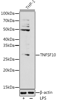
Western blot analysis of lysates from THP-1 cells, using (A2138) at 1:1000 dilution. THP-1 cells were treated with LPS (1 µg/ml) at 37°C for 8 hours. Secondary antibody: HRP-conjugated Goat anti-Rabbit IgG (H+L) (AS014) at 1:10000 dilution. Lysates/proteins: 25µg per lane. Blocking buffer: 3% nonfat dry milk in TBST. Detection: ECL Basic Kit (RM00020). Exposure time: 180s. |
Produktgarantie und fachkundiger Support
