
CDKN1A/p21CIP1 Rabbit mAb, Unconjugated, Monoclonal
Artikelnummer:
ABB-A21897
- Bilder (8)
| Artikelname: | CDKN1A/p21CIP1 Rabbit mAb, Unconjugated, Monoclonal |
| Artikelnummer: | ABB-A21897 |
| Hersteller Artikelnummer: | A21897 |
| Alternativnummer: | ABB-A21897-20UL,ABB-A21897-100UL,ABB-A21897-500UL,ABB-A21897-1000UL |
| Hersteller: | ABclonal |
| Wirt: | Rabbit |
| Kategorie: | Antikörper |
| Applikation: | ELISA, IHC-P, WB |
| Spezies Reaktivität: | Human |
| Immunogen: | Recombinant protein (or fragment).This information is considered to be commercially sensitive. |
| Konjugation: | Unconjugated |
| Alternative Synonym: | P21, CIP1, SDI1, WAF1, CAP20, CDKN1, MDA-6, p21CIP1, CDKN1A/p21CIP1 |
| This gene encodes a potent cyclin-dependent kinase inhibitor. The encoded protein binds to and inhibits the activity of cyclin-cyclin-dependent kinase2 or -cyclin-dependent kinase4 complexes, and thus functions as a regulator of cell cycle progression at G1. The expression of this gene is tightly controlled by the tumor suppressor protein p53, through which this protein mediates the p53-dependent cell cycle G1 phase arrest in response to a variety of stress stimuli. This protein can interact with proliferating cell nuclear antigen, a DNA polymerase accessory factor, and plays a regulatory role in S phase DNA replication and DNA damage repair. This protein was reported to be specifically cleaved by CASP3-like caspases, which thus leads to a dramatic activation of cyclin-dependent kinase2, and may be instrumental in the execution of apoptosis following caspase activation. Mice that lack this gene have the ability to regenerate damaged or missing tissue. Multiple alternatively spliced variants have been found for this gene. |
| Klonalität: | Monoclonal |
| Klon-Bezeichnung: | [ARC51046] |
| Molekulargewicht: | 18kDa |
| NCBI: | 1026 |
| UniProt: | P38936 |
| Reinheit: | Affinity purification |
| Sequenz: | MSEPAGDVRQNPCGSKACRRLFGPVDSEQLSRDCDALMAGCIQEARERWNFDFVTETPLEGDFAWERVRGLGLPKLYLPTGPRRGRDELGGGRRPGTSPALLQGTAEEDHVDLSLSCTLVPRSGEQAEGSPGGPGDSQGRKRRQTSMTDFYHSKRRLIFSKRKP |
| Target-Kategorie: | CDKN1A |
| Antibody Type: | Primary Antibody |
| Application Verdünnung: | WB,1:500 - 1:1000|IHC-P,1:500 - 1:1000|ELISA,Recommended starting concentration is 1 µg/mL. Please optimize the concentration based on your specific assay requirements. |
| Anwendungsbeschreibung: | Cross-Reactivity: Human. ResearchArea: Epigenetics Nuclear Signaling,DNA Damage Repair,Cancer,Tumor suppressors,p53 pathway,Signal Transduction,Kinase,PI3K-Akt Signaling Pathway,ATM Signaling Pathway,Cell Biology Developmental Biology,Apoptosis,Cell Cycle,Cell cycle inhibitors,Cell Cycle Control-G1 S Checkpoint,Cell Cycle Control-G2 M DNA Damage Checkpoint,Endocrine Metabolism,AMPK Signaling Pathway,Immunology Inflammation,Stem Cells. Shipping: Ice Bag |
|
|
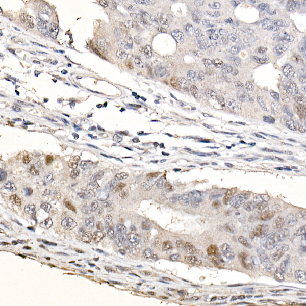
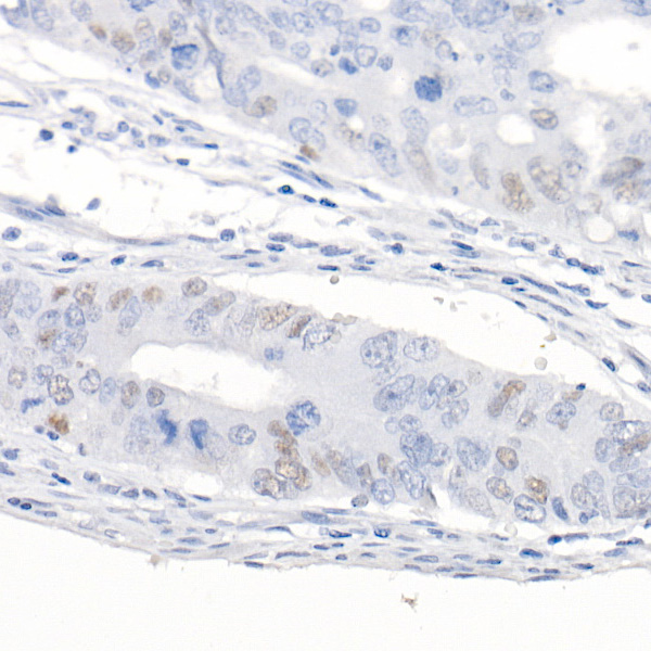
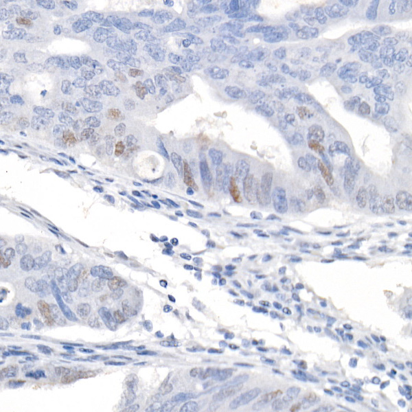
Immunohistochemistry analysis of paraffin-embedded Human colon cancer (100 times dilution of primary antibody) using CDKN1A/p21CIP1 Rabbit mAb (A21897) at dilution of 1:1000 (40x lens). High pressure antigen retrieval performed with 0.01M Citrate buffer (pH 6.0) prior to IHC staining. |
|
|
Immunohistochemistry analysis of paraffin-embedded Human colon cancer (1600 times dilution of primary antibody) using CDKN1A/p21CIP1 Rabbit mAb (A21897) at dilution of 1:1000 (40x lens). High pressure antigen retrieval performed with 0.01M Citrate buffer (pH 6.0) prior to IHC staining. |
|
|
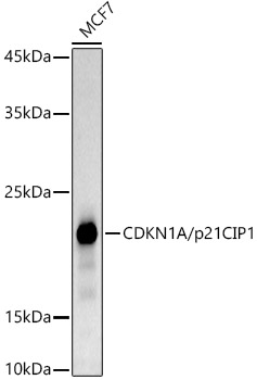
Western blot analysis of lysates from MCF7 cells, using CDKN1A/p21CIP1 Rabbit mAb (A21897) at1:1000 dilution. Secondary antibody: HRP-conjugated Goat anti-Rabbit IgG (H+L) (AS014) at 1:10000 dilution. Lysates/proteins: 25µg per lane. Blocking buffer: 3% nonfat dry milk in TBST. Detection: ECL Basic Kit (RM00020). Exposure time: 180s. |
|
|
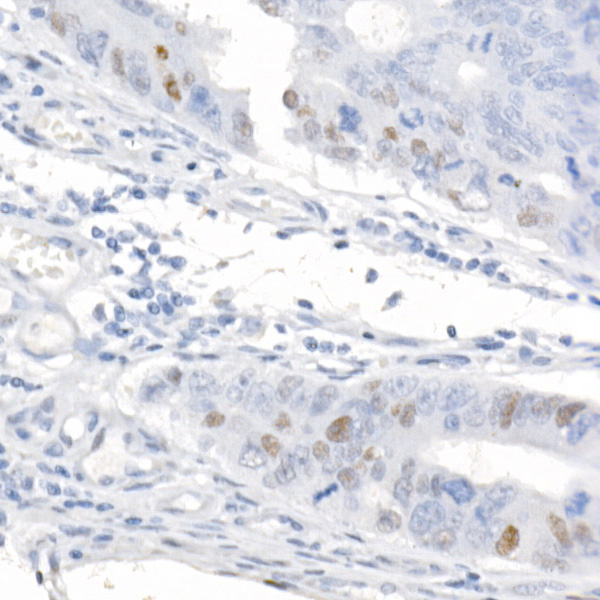
Immunohistochemistry analysis of paraffin-embedded Human colon cancer (200 times dilution of primary antibody) using CDKN1A/p21CIP1 Rabbit mAb (A21897) at dilution of 1:1000 (40x lens). High pressure antigen retrieval performed with 0.01M Citrate buffer (pH 6.0) prior to IHC staining. |
|
|
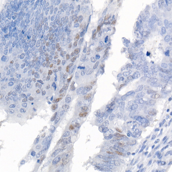
Immunohistochemistry analysis of paraffin-embedded Human colon cancer (400 times dilution of primary antibody) using CDKN1A/p21CIP1 Rabbit mAb (A21897) at dilution of 1:1000 (40x lens). High pressure antigen retrieval performed with 0.01M Citrate buffer (pH 6.0) prior to IHC staining. |
|
|
Immunohistochemistry analysis of paraffin-embedded Human colon cancer (800 times dilution of primary antibody) using CDKN1A/p21CIP1 Rabbit mAb (A21897) at dilution of 1:1000 (40x lens). High pressure antigen retrieval performed with 0.01M Citrate buffer (pH 6.0) prior to IHC staining. |
|
|
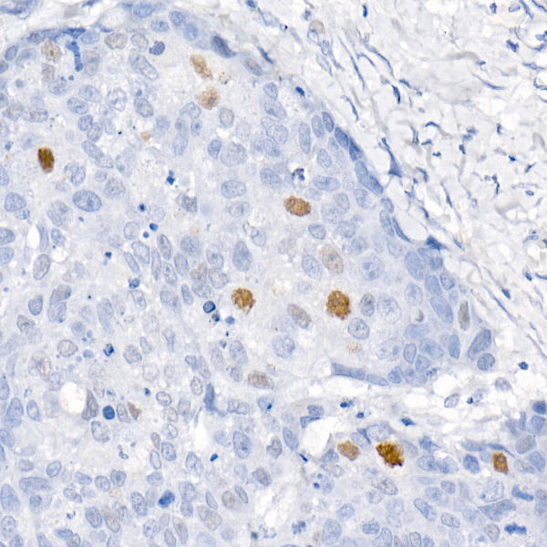
Immunohistochemistry analysis of paraffin-embedded Human colon cancer (positive control antibody staining) using CDKN1A/p21CIP1 Rabbit mAb (A21897) at dilution of 1:1000 (40x lens). High pressure antigen retrieval performed with 0.01M Citrate buffer (pH 6.0) prior to IHC staining. |
|
|
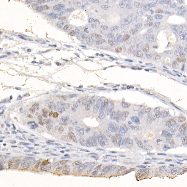
Immunohistochemistry analysis of paraffin-embedded Human esophageal cancer using CDKN1A/p21CIP1 Rabbit mAb (A21897) at dilution of 1:1000 (40x lens). High pressure antigen retrieval performed with 0.01M Citrate buffer (pH 6.0) prior to IHC staining. |
Produktgarantie und fachkundiger Support
