
MonoMethyl-Histone H3-K4 Rabbit mAb, Unconjugated
Artikelnummer:
ABB-A22078
- Bilder (8)

| Artikelname: | MonoMethyl-Histone H3-K4 Rabbit mAb, Unconjugated |
| Artikelnummer: | ABB-A22078 |
| Hersteller Artikelnummer: | A22078 |
| Alternativnummer: | ABB-A22078-20UL,ABB-A22078-100UL,ABB-A22078-500UL,ABB-A22078-1000UL |
| Hersteller: | ABclonal |
| Wirt: | Rabbit |
| Kategorie: | Antikörper |
| Applikation: | CUT&Tag, DOT, ELISA, IF, WB |
| Spezies Reaktivität: | Human |
| Immunogen: | Synthetic peptide. This information is considered to be commercially sensitive. |
| Konjugation: | Unconjugated |
| Alternative Synonym: | H3t, H3.4, H3/g, H3FT, H3C16, HIST3H3, MonoMethyl-Histone H3-K4 |
| Histones are basic nuclear proteins that are responsible for the nucleosome structure of the chromosomal fiber in eukaryotes. Nucleosomes consist of approximately 146 bp of DNA wrapped around a histone octamer composed of pairs of each of the four core histones (H2A, H2B, H3, and H4). The chromatin fiber is further compacted through the interaction of a linker histone, H1, with the DNA between the nucleosomes to form higher order chromatin structures. This gene is intronless and encodes a replication-dependent histone that is a member of the histone H3 family. Transcripts from this gene lack polyA tails, instead, they contain a palindromic termination element. This gene is located separately from the other H3 genes that are in the histone gene cluster on chromosome 6p22-p21.3. |
| Klonalität: | Monoclonal |
| Klon-Bezeichnung: | [ARC54646] |
| Molekulargewicht: | 15 kDa |
| NCBI: | 8290 |
| UniProt: | Q16695 |
| Reinheit: | Affinity purification |
| Sequenz: | MARTKQTARKSTGGKAPRKQLATKAARKSAPATGGVKKPHRYRPGTVALREIRRYQKSTELLIRKLPFQRLVREIAQDFKTDLRFQSSAVMALQEACEAY |
| Target-Kategorie: | H3C1/H3-4 |
| Antibody Type: | Primary Antibody |
| Application Verdünnung: | WB,1:1000 - 1:20000|DB,1:1000 - 1:5000|IF/ICC,1:100 - 1:500|ELISA,Recommended starting concentration is 1 µg/mL. Please optimize the concentration based on your specific assay requirements.|CUT&Tag, 105 cells /1 µg |
| Anwendungsbeschreibung: | Cross-Reactivity: Human,Mouse,Rat,Other (Wide Range Predicted). ResearchArea: Epigenetics Nuclear Signaling,Protein phosphorylation,Signal Transduction,MAPK-Erk Signaling Pathway. Shipping: Ice Bag |
|
|
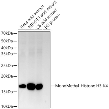
Western blot analysis of various lysates using MonoMethyl-Histone H3-K4 Rabbit mAb (A22078) at 1:1000 dilution incubated overnight at 4°C. Secondary antibody: HRP-conjugated Goat anti-Rabbit IgG (H+L) (AS014) at 1:10000 dilution. Lysates/proteins: 25 µg per lane. Blocking buffer: 3% nonfat dry milk in TBST. Detection: ECL Basic Kit (RM00020). Exposure time: 45s. |
|
|
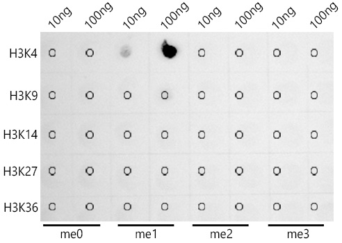
Dot-blot analysis of all sorts of peptides using MonoMethyl-Histone H3-K4 antibody (A22078) at 1:2000 dilution. |
|
|
Western blot analysis of various lysates using MonoMethyl-Histone H3-K4 Rabbit mAb (A22078) at1:20000 dilution. Secondary antibody: HRP-conjugated Goat anti-Rabbit IgG (H+L) (AS014) at 1:10000 dilution. Lysates/proteins: 25µg per lane. Blocking buffer: 3% nonfat dry milk in TBST. Detection: ECL Enhanced Kit (RM00021). Exposure time: 60s. |
|
|
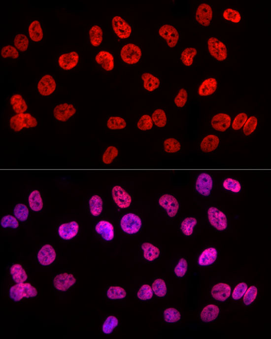
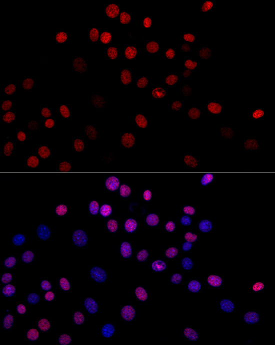
Immunofluorescence analysis of HeLa cells using MonoMethyl-Histone H3-K4 Rabbit mAb (A22078) at dilution of 1:300 (40x lens). Secondary antibody: Cy3-conjugated Goat anti-Rabbit IgG (H+L) (AS007) at 1:500 dilution. Blue: DAPI for nuclear staining. |
|
|
Immunofluorescence analysis of NIH/3T3 cells using MonoMethyl-Histone H3-K4 Rabbit mAb (A22078) at dilution of 1:300 (40x lens). Secondary antibody: Cy3-conjugated Goat anti-Rabbit IgG (H+L) (AS007) at 1:500 dilution. Blue: DAPI for nuclear staining. |
|
|
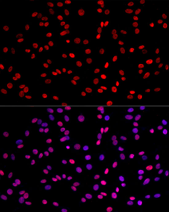
Immunofluorescence analysis of PC-12 cells using MonoMethyl-Histone H3-K4 Rabbit mAb (A22078) at dilution of 1:300 (40x lens). Secondary antibody: Cy3-conjugated Goat anti-Rabbit IgG (H+L) (AS007) at 1:500 dilution. Blue: DAPI for nuclear staining. |
|
|
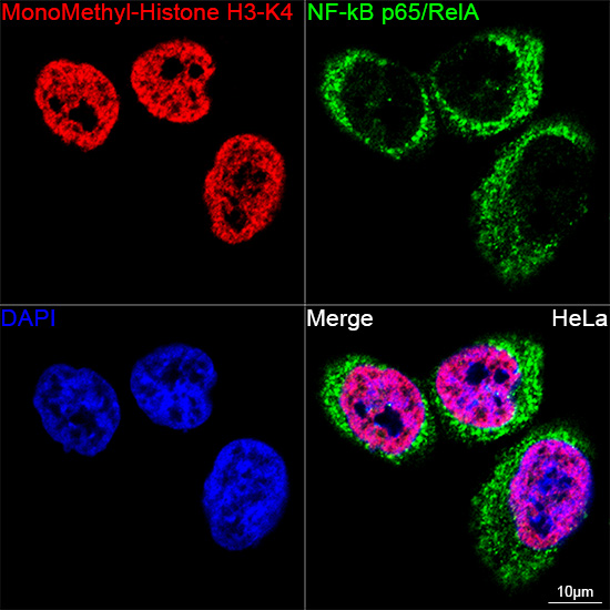
Confocal imaging of HeLa cells using MonoMethyl-Histone H3-K4RabbitmAb (A22078, dilution 1:300) (Green). The cells were counterstained with [KOValidated]NF-kBp65/RelA Rabbit mAb (A22331, dilution 1:100) (Red). DAPI was used for nuclear staining (blue). Objective: 60x. |
|
|
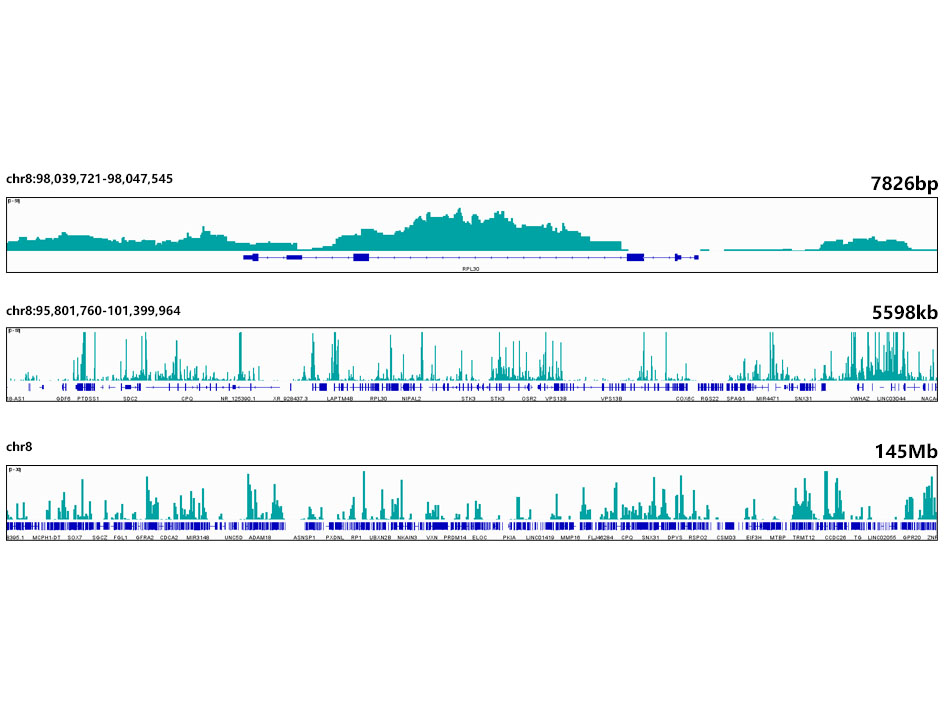
CUT&,Tag was performed using the CUT&,Tag Assay Kit (pAG-Tn5) for Illumina(RK20265) from 105 K562 cells with 1 µg MonoMethyl-Histone H3-K4 antibody (A22078), along with a Goat Anti-Rabbit IgG(H+L). The CUT&,Tag results indicate the enrichment pattern of H3K4me1 in representative gene loci (RPL30), as shown in figure. |
Produktgarantie und fachkundiger Support
