
PHGDH Rabbit mAb, Unconjugated, Monoclonal
Artikelnummer:
ABB-A22129
- Bilder (8)

| Artikelname: | PHGDH Rabbit mAb, Unconjugated, Monoclonal |
| Artikelnummer: | ABB-A22129 |
| Hersteller Artikelnummer: | A22129 |
| Alternativnummer: | ABB-A22129-100UL,ABB-A22129-20UL |
| Hersteller: | ABclonal |
| Wirt: | Rabbit |
| Kategorie: | Antikörper |
| Applikation: | ELISA, IF, IHC-P, WB |
| Spezies Reaktivität: | Human |
| Immunogen: | Recombinant protein (or fragment).This information is considered to be commercially sensitive. |
| Konjugation: | Unconjugated |
| Alternative Synonym: | NLS, PDG, PGD, NLS1, PGAD, PGDH, SERA, 3PGDH, 3-PGDH, PHGDHD, HEL-S-113, PHGDH |
| This gene encodes the enzyme which is involved in the early steps of L-serine synthesis in animal cells. L-serine is required for D-serine and other amino acid synthesis. The enzyme requires NAD/NADH as a cofactor and forms homotetramers for activity. Mutations in this gene have been found in a family with congenital microcephaly, psychomotor retardation and other symptoms. Multiple alternatively spliced transcript variants have been found, however the full-length nature of most are not known. |
| Application Verdünnung: | WB,1:5000 - 1:20000|IHC-P,1:2000 - 1:8000|IF/ICC,1:400 - 1:1600|ELISA,Recommended starting concentration is 1 µg/mL. Please optimize the concentration based on your specific assay requirements. |
| Anwendungsbeschreibung: | Cross-Reactivity: Human,Mouse,Rat. ResearchArea: Signal Transduction,Endocrine Metabolism,Amino acid metabolism,Neuroscience. Shipping: Ice Bag |
|
|
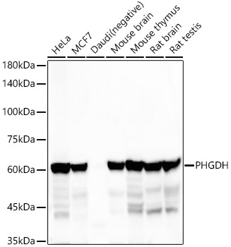
Western blot analysis of various lysates using PHGDH Rabbit mAb (A22129) at 1:20000 dilution incubated at room temperature for 1.5 hours. Secondary antibody: HRP-conjugated Goat anti-Rabbit IgG (H+L) (AS014) at 1:10000 dilution. Lysates/proteins: 25 µg per lane. Blocking buffer: 3% nonfat dry milk in TBST. Detection: ECL Basic Kit (RM00020). Exposure time: 45s. |
|
|
Western blot analysis of lysates from Rat brain using PHGDH Rabbit mAb (A22129) at 1:20000 dilution incubated at room temperature for 1.5 hours. Secondary antibody: HRP-conjugated Goat anti-Rabbit IgG (H+L) (AS014) at 1:10000 dilution. Lysates/proteins: 25 µg per lane. Blocking buffer: 3% nonfat dry milk in TBST. Detection: ECL Basic Kit (RM00020). Exposure time: 90s. |
|
|
Immunohistochemistry analysis of paraffin-embedded Human esophagus tissue using PHGDH Rabbit mAb (A22129) at a dilution of 1:2500 (40x lens). High pressure antigen retrieval performed with 0.01M Tris-EDTA Buffer (pH 9.0) prior to IHC staining. |
|
|
Immunohistochemistry analysis of paraffin-embedded Human colon cancer tissue using PHGDH Rabbit mAb (A22129) at a dilution of 1:2500 (40x lens). High pressure antigen retrieval performed with 0.01M Tris-EDTA Buffer (pH 9.0) prior to IHC staining. |
|
|
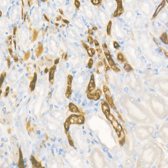
Immunohistochemistry analysis of paraffin-embedded Human kidney tissue using PHGDH Rabbit mAb (A22129) at a dilution of 1:2500 (40x lens). High pressure antigen retrieval performed with 0.01M Tris-EDTA Buffer (pH 9.0) prior to IHC staining. |
|
|
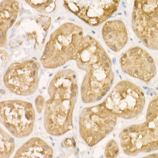
Immunohistochemistry analysis of paraffin-embedded Mouse kidney tissue using PHGDH Rabbit mAb (A22129) at a dilution of 1:2500 (40x lens). High pressure antigen retrieval performed with 0.01M Tris-EDTA Buffer (pH 9.0) prior to IHC staining. |
|
|
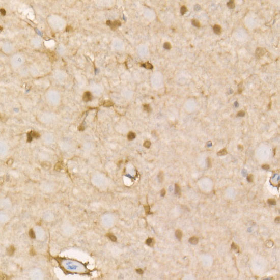
Immunohistochemistry analysis of paraffin-embedded Rat brain tissue using PHGDH Rabbit mAb (A22129) at a dilution of 1:2500 (40x lens). High pressure antigen retrieval performed with 0.01M Tris-EDTA Buffer (pH 9.0) prior to IHC staining. |
|
|
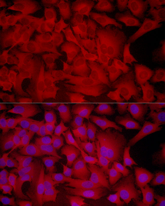
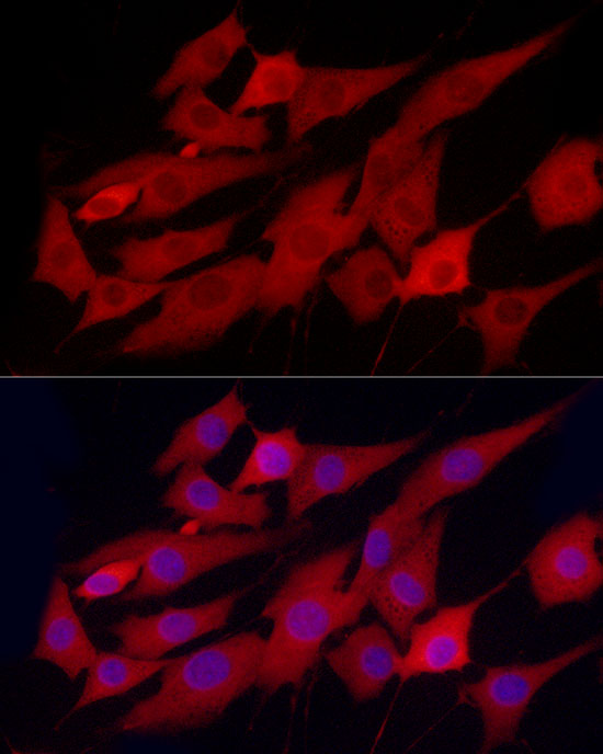
Confocal imaging of HeLa cells using PHGDH Rabbit mAb (A22129, dilution 1:1000) followed by a further incubation with Cy3 Goat Anti-Rabbit IgG (H+L) (AS007, dilution 1:500) (Red). The cells were counterstained with alpha-Tubulin Mouse mAb (AC012, dilution 1:400) followed by incubation with ABflo 488-conjugated Goat Anti-Mouse IgG (H+L) Ab (AS076, dilution 1:500) (Green). DAPI was used for nuclear staining (Blue). Objective: 100x. |
Produktgarantie und fachkundiger Support
