
PGAM5 Rabbit mAb, Unconjugated, Monoclonal
Artikelnummer:
ABB-A22203
- Bilder (8)

| Artikelname: | PGAM5 Rabbit mAb, Unconjugated, Monoclonal |
| Artikelnummer: | ABB-A22203 |
| Hersteller Artikelnummer: | A22203 |
| Alternativnummer: | ABB-A22203-100UL,ABB-A22203-20UL |
| Hersteller: | ABclonal |
| Wirt: | Rabbit |
| Kategorie: | Antikörper |
| Applikation: | ELISA, IF, IHC-P, WB |
| Spezies Reaktivität: | Human |
| Immunogen: | Recombinant protein (or fragment).This information is considered to be commercially sensitive. |
| Konjugation: | Unconjugated |
| Alternative Synonym: | BXLBV68, PGAM5 |
| Enables GTPase activator activity and protein serine/threonine phosphatase activity. Involved in necroptotic process. Located in mitochondrion. |
| Application Verdünnung: | WB,1:20000 - 1:80000|IHC-P,1:100 - 1:500|IF/ICC,1:300 - 1:1200|ELISA,Recommended starting concentration is 1 µg/mL. Please optimize the concentration based on your specific assay requirements. |
| Anwendungsbeschreibung: | Cross-Reactivity: Human. ResearchArea: Signal Transduction. Shipping: Ice Bag |
|
|
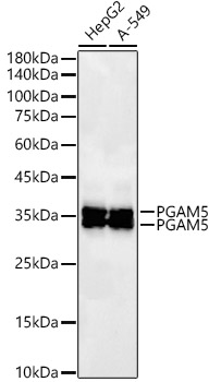
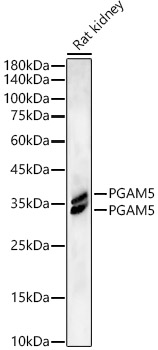
Western blot analysis of various lysates, using PGAM5 Rabbit mAb (A22203) at1:20000 dilution. Secondary antibody: HRP-conjugated Goat anti-Rabbit IgG (H+L) (AS014) at 1:10000 dilution. Lysates/proteins: 25µg per lane. Blocking buffer: 3% nonfat dry milk in TBST. Detection: ECL Basic Kit (RM00020). Exposure time: 30s. |
|
|
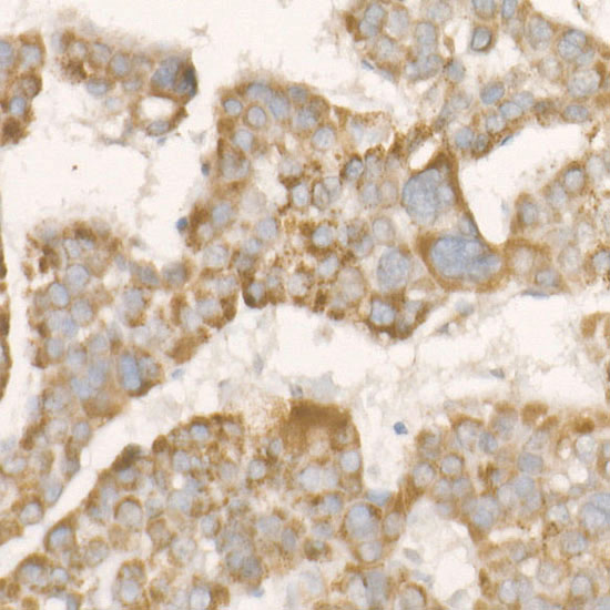
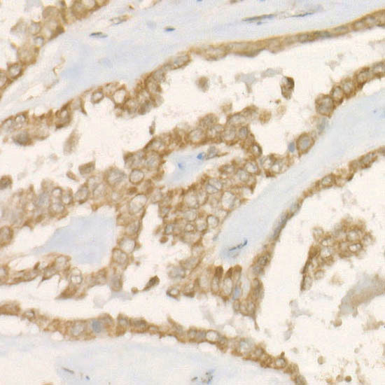
Immunohistochemistry analysis of paraffin-embedded Human esophagus tissue using PGAM5 Rabbit mAb (A22203) at a dilution of 1:500 (40x lens). High pressure antigen retrieval performed with 0.01M Tris-EDTA Buffer (pH 9.0) prior to IHC staining. |
|
|
Immunohistochemistry analysis of paraffin-embedded Human breast cancer tissue using PGAM5 Rabbit mAb (A22203) at a dilution of 1:500 (40x lens). High pressure antigen retrieval performed with 0.01M Tris-EDTA Buffer (pH 9.0) prior to IHC staining. |
|
|
Immunohistochemistry analysis of paraffin-embedded Human thyroid cancer tissue using PGAM5 Rabbit mAb (A22203) at a dilution of 1:500 (40x lens). High pressure antigen retrieval performed with 0.01M Tris-EDTA Buffer (pH 9.0) prior to IHC staining. |
|
|
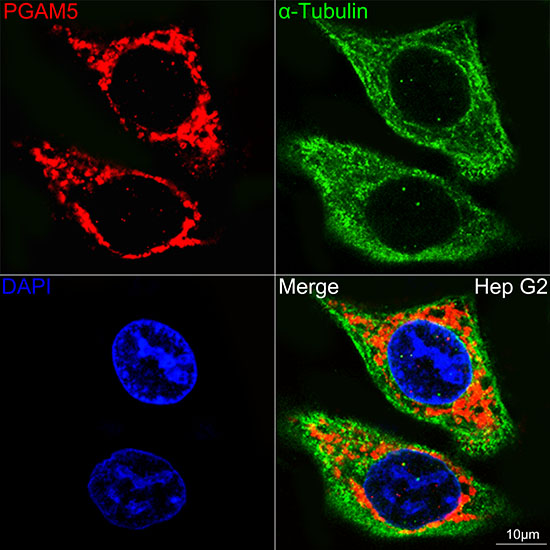
Confocal imaging of Hep G2 cells using PGAM5 Rabbit mAb (A22203,dilution 1:300)(Red). The cells were counterstained with alpha-Tubulin Mouse mAb (AC012,dilution 1:400) (Green). DAPI was used for nuclear staining (blue). Objective: 60x. |
Produktgarantie und fachkundiger Support
