
HCoV-229E Spike S1 Rabbit mAb, Unconjugated, Monoclonal
Artikelnummer:
ABB-A22605
- Bilder (1)
| Artikelname: | HCoV-229E Spike S1 Rabbit mAb, Unconjugated, Monoclonal |
| Artikelnummer: | ABB-A22605 |
| Hersteller Artikelnummer: | A22605 |
| Alternativnummer: | ABB-A22605-100UL,ABB-A22605-20UL |
| Hersteller: | ABclonal |
| Wirt: | Rabbit |
| Kategorie: | Antikörper |
| Applikation: | ELISA, WB |
| Immunogen: | Recombinant protein (or fragment).This information is considered to be commercially sensitive. |
| Konjugation: | Unconjugated |
| S1 region attaches the virion to the cell membrane by interacting with host ANPEP/aminopeptidase N, initiating the infection. Binding to the receptor probably induces conformational changes in the S glycoprotein unmasking the fusion peptide of S2 region and activating membranes fusion. S2 region belongs to the class I viral fusion protein. Under the current model, the protein has at least 3 conformational states: pre-fusion native state, pre-hairpin intermediate state, and post-fusion hairpin state. During viral and target cell membrane fusion, the coiled coil regions (heptad repeats regions assume a trimer-of-hairpins structure, positioning the fusion peptide in close proximity to the C-terminal region of the ectodomain. The formation of this structure appears to drive apposition and subsequent fusion of viral and target cell membranes. |
| Application Verdünnung: | WB,1:500 - 1:1000|ELISA,Recommended starting concentration is 1 µg/mL. Please optimize the concentration based on your specific assay requirements. |
| Anwendungsbeschreibung: | Cross-Reactivity: HCoV-229E. Shipping: Ice Bag |
|
|
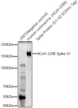
Western blot analysis of various lysates, using HCoV-229E Spike S1 Rabbit mAb (A22605) at 1:800 dilution. Secondary antibody: HRP-conjugated Goat anti-Rabbit IgG (H+L) (AS014) at 1:10000 dilution. Lysates/proteins: 25µg per lane. Blocking buffer: 3% nonfat dry milk in TBST. Detection: ECL Enhanced Kit (RM00021). Negative control (NC): 293T. Exposure time: 180s. |
Produktgarantie und fachkundiger Support
