
HSF1 Rabbit mAb, Unconjugated, Monoclonal
Artikelnummer:
ABB-A23895
- Bilder (8)

| Artikelname: | HSF1 Rabbit mAb, Unconjugated, Monoclonal |
| Artikelnummer: | ABB-A23895 |
| Hersteller Artikelnummer: | A23895 |
| Alternativnummer: | ABB-A23895-100UL,ABB-A23895-20UL |
| Hersteller: | ABclonal |
| Wirt: | Rabbit |
| Kategorie: | Antikörper |
| Applikation: | ELISA, IHC-P, IP, WB |
| Spezies Reaktivität: | Human |
| Immunogen: | Synthetic peptide. This information is considered to be commercially sensitive. |
| Konjugation: | Unconjugated |
| Alternative Synonym: | HSTF1, HSF1 |
| The product of this gene is a transcription factor that is rapidly induced after temperature stress and binds heat shock promoter elements (HSE). This protein plays a role in the regulation of lifespan. Expression of this gene is repressed by phosphorylation, which promotes binding by heat shock protein 90. |
| Application Verdünnung: | WB,1:1000 - 1:4000|IHC-P,1:200 - 1:2000|IP,0.5µg-4µg antibody for 200µg-400µg extracts of whole cells|ELISA,Recommended starting concentration is 1 µg/mL. Please optimize the concentration based on your specific assay requirements. |
| Anwendungsbeschreibung: | Cross-Reactivity: Human,Mouse,Rat. ResearchArea: Epigenetics Nuclear Signaling,Transcription Factors,Signal Transduction,MAPK-JNK Signaling Pathway,Cardiovascular,Heart,Cardiogenesis. Shipping: Ice Bag |
|
|
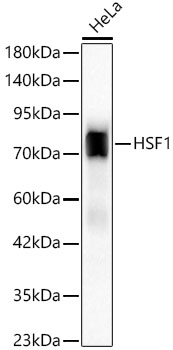
Western blot analysis of lysates from HeLa cells using HSF1 Rabbit mAb (A23895) at 1:1000 dilution incubated overnight at 4°C. Secondary antibody: HRP-conjugated Goat anti-Rabbit IgG (H+L) (AS014) at 1:10000 dilution. Lysates/proteins: 25 µg per lane. Blocking buffer: 3% nonfat dry milk in TBST. Detection: ECL Basic Kit (RM00020). Exposure time: 30s. |
|
|
Western blot analysis of lysates from Rat testis using HSF1 Rabbit mAb (A23895) at 1:1000 dilution incubated overnight at 4°C. Secondary antibody: HRP-conjugated Goat anti-Rabbit IgG (H+L) (AS014) at 1:10000 dilution. Lysates/proteins: 25 µg per lane. Blocking buffer: 3% nonfat dry milk in TBST. Detection: ECL Basic Kit (RM00020). Exposure time: 90s. |
|
|
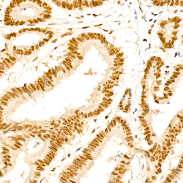
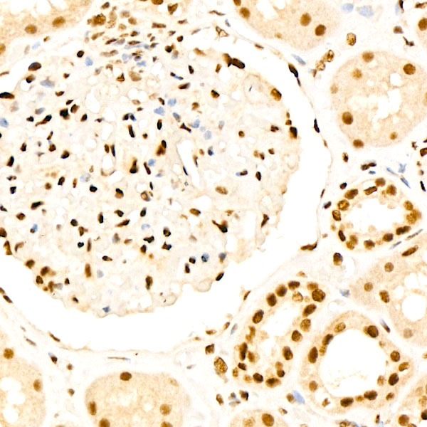
Immunohistochemistry analysis of paraffin-embedded Human colon tissue using HSF1 Rabbit mAb (A23895) at a dilution of 1:1000 (40x lens). High pressure antigen retrieval performed with 0.01M Tris-EDTA Buffer (pH 9.0) prior to IHC staining. |
|
|
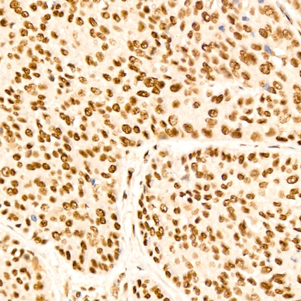
Immunohistochemistry analysis of paraffin-embedded Human breast cancer tissue using HSF1 Rabbit mAb (A23895) at a dilution of 1:1000 (40x lens). High pressure antigen retrieval performed with 0.01M Tris-EDTA Buffer (pH 9.0) prior to IHC staining. |
|
|
Immunohistochemistry analysis of paraffin-embedded Human thyroid cancer tissue using HSF1 Rabbit mAb (A23895) at a dilution of 1:1000 (40x lens). High pressure antigen retrieval performed with 0.01M Tris-EDTA Buffer (pH 9.0) prior to IHC staining. |
|
|
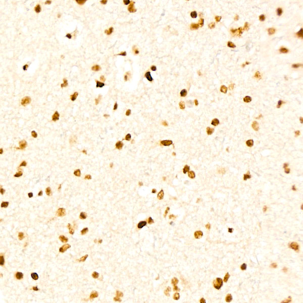
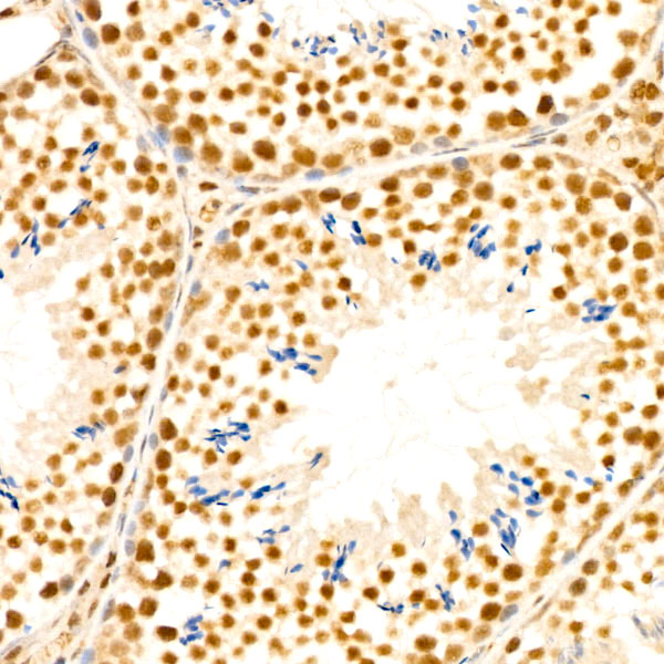
Immunohistochemistry analysis of paraffin-embedded Mouse testis tissue using HSF1 Rabbit mAb (A23895) at a dilution of 1:1000 (40x lens). High pressure antigen retrieval performed with 0.01M Tris-EDTA Buffer (pH 9.0) prior to IHC staining. |
|
|
Immunohistochemistry analysis of paraffin-embedded Rat colon tissue using HSF1 Rabbit mAb (A23895) at a dilution of 1:1000 (40x lens). High pressure antigen retrieval performed with 0.01M Tris-EDTA Buffer (pH 9.0) prior to IHC staining. |
|
|
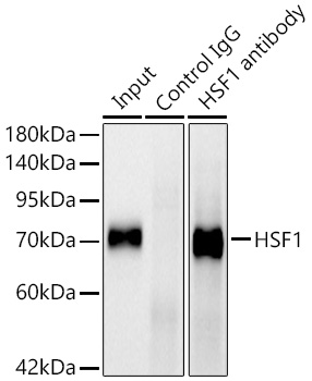
Immunoprecipitation analysis of 300 µg extracts of HeLa cells using 3 µg HSF1 Rabbit mAb (A23895). Western blot was performed from the immunoprecipitate using HSF1 Rabbit mAb (A23895) at a dilition of 1:2000. |
Produktgarantie und fachkundiger Support
