
NPC2 Rabbit pAb, Unconjugated, Polyclonal
Artikelnummer:
ABB-A5413
- Bilder (8)


| Artikelname: | NPC2 Rabbit pAb, Unconjugated, Polyclonal |
| Artikelnummer: | ABB-A5413 |
| Hersteller Artikelnummer: | A5413 |
| Alternativnummer: | ABB-A5413-100UL,ABB-A5413-20UL,ABB-A5413-1000UL,ABB-A5413-500UL |
| Hersteller: | ABclonal |
| Wirt: | Rabbit |
| Kategorie: | Antikörper |
| Applikation: | ELISA, IF, IHC-P, WB |
| Spezies Reaktivität: | Human |
| Immunogen: | Recombinant protein (or fragment).This information is considered to be commercially sensitive. |
| Konjugation: | Unconjugated |
| Alternative Synonym: | HE1, EDDM1, NPC2 |
| This gene encodes a protein containing a lipid recognition domain. The encoded protein may function in regulating the transport of cholesterol through the late endosomal/lysosomal system. Mutations in this gene have been associated with Niemann-Pick disease, type C2 and frontal lobe atrophy. |
| Klonalität: | Polyclonal |
| Molekulargewicht: | 17kDa |
| NCBI: | 10577 |
| UniProt: | P61916 |
| Reinheit: | Affinity purification |
| Sequenz: | EPVQFKDCGSVDGVIKEVNVSPCPTQPCQLSKGQSYSVNVTFTSNIQSKSSKAVVHGILMGVPVPFPIPEPDGCKSGINCPIQKDKTYSYLNKLPVKSEYPSIKLVVEWQLQDDKNQSLFCWEIPVQIVSHL |
| Target-Kategorie: | NPC2 |
| Antibody Type: | Primary Antibody |
| Application Verdünnung: | WB,1:500 - 1:2000|IF-P,1:50 - 1:200|IHC-P,1:50 - 1:200|ELISA,Recommended starting concentration is 1 µg/mL. Please optimize the concentration based on your specific assay requirements. |
| Anwendungsbeschreibung: | Cross-Reactivity: Human,Mouse,Rat. ResearchArea: Signal Transduction,Endocrine Metabolism,Lipid Metabolism,Cholesterol Metabolism,Neuroscience,Neurodegenerative Diseases,Cardiovascular,Lipids. Shipping: Ice Bag |
|
|
Western blot analysis of lysates from wild type (WT) and NPC2 knockdown (KD) 293T cells using NPC2 Rabbit pAb (A5413) at 1:1000 dilution incubated overnight at 4°C. Secondary antibody: HRP-conjugated Goat anti-Rabbit IgG (H+L) (AS014) at 1:10000 dilution. Lysates/proteins: 25 µg per lane. Blocking buffer: 3% nonfat dry milk in TBST. Detection: ECL Basic Kit (RM00020). Exposure time: 45s. |
|
|
Western blot analysis of lysates from Hep G2 cells using NPC2 Rabbit pAb (A5413) at 1:1000 dilution incubated overnight at 4°C. Secondary antibody: HRP-conjugated Goat anti-Rabbit IgG (H+L) (AS014) at 1:10000 dilution. Lysates/proteins: 25 µg per lane. Blocking buffer: 3% nonfat dry milk in TBST. Detection: ECL Basic Kit (RM00020). Exposure time: 45s. |
|
|
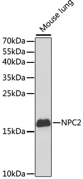
Western blot analysis of lysates from mouse lung, using NPC2 Rabbit pAb (A5413) at 1:1000 dilution. Secondary antibody: HRP-conjugated Goat anti-Rabbit IgG (H+L) (AS014) at 1:10000 dilution. Lysates/proteins: 25µg per lane. Blocking buffer: 3% nonfat dry milk in TBST. Detection: ECL Basic Kit (RM00020). Exposure time: 90s. |
|
|
Western blot analysis of lysates from NIH/3T3 cells using NPC2 Rabbit pAb (A5413) at 1:1000 dilution incubated overnight at 4°C. Secondary antibody: HRP-conjugated Goat anti-Rabbit IgG (H+L) (AS014) at 1:10000 dilution. Lysates/proteins: 25 µg per lane. Blocking buffer: 3% nonfat dry milk in TBST. Detection: ECL Basic Kit (RM00020). Exposure time: 30s. |
|
|
Immunohistochemistry analysis of paraffin-embedded Mouse testis using NPC2 Rabbit pAb (A5413) at dilution of 1:50 (40x lens). High pressure antigen retrieval performed with 0.01M Citrate buffer (pH 6.0) prior to IHC staining. |
|
|
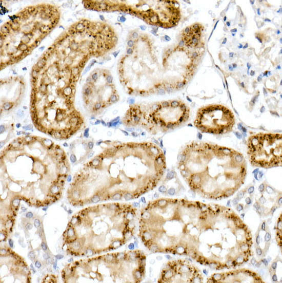
Immunohistochemistry analysis of paraffin-embedded Rat kidney using NPC2 Rabbit pAb (A5413) at dilution of 1:50 (40x lens). High pressure antigen retrieval performed with 0.01M Citrate buffer (pH 6.0) prior to IHC staining. |
|
|
Immunofluorescence analysis of Human liver tissue using NPC2 Rabbit pAb (A5413) at a dilution of 1:100 (40x lens). Secondary antibody: Cy3-conjugated Goat anti-Rabbit IgG (H+L)(AS007) at 1:500 dilution. Blue: DAPI for nuclear staining. High pressure antigen retrieval performed with 0.01M Citrate Buffer (pH 6.0) prior to IF staining. |
|
|
Immunofluorescence analysis of Mouse lung tissue using NPC2 Rabbit pAb (A5413) at a dilution of 1:100 (40x lens). Secondary antibody: Cy3-conjugated Goat anti-Rabbit IgG (H+L)(AS007) at 1:500 dilution. Blue: DAPI for nuclear staining. High pressure antigen retrieval performed with 0.01M Citrate Buffer (pH 6.0) prior to IF staining. |
Produktgarantie und fachkundiger Support
