
PGRMC1 Rabbit pAb, Unconjugated, Polyclonal
Artikelnummer:
ABB-A5619
- Bilder (8)
| Artikelname: | PGRMC1 Rabbit pAb, Unconjugated, Polyclonal |
| Artikelnummer: | ABB-A5619 |
| Hersteller Artikelnummer: | A5619 |
| Alternativnummer: | ABB-A5619-100UL,ABB-A5619-20UL,ABB-A5619-500UL,ABB-A5619-1000UL |
| Hersteller: | ABclonal |
| Wirt: | Rabbit |
| Kategorie: | Antikörper |
| Applikation: | ELISA, IF, IHC-P, WB |
| Spezies Reaktivität: | Human |
| Immunogen: | Recombinant protein (or fragment).This information is considered to be commercially sensitive. |
| Konjugation: | Unconjugated |
| Alternative Synonym: | IZA, MPR, Dap1, HPR6.6, PGRMC1 |
| This gene encodes a putative membrane-associated progesterone steroid receptor. The protein is expressed predominantly in the liver and kidney. |
| Klonalität: | Polyclonal |
| Molekulargewicht: | 22kDa |
| NCBI: | 10857 |
| UniProt: | O00264 |
| Reinheit: | Affinity purification |
| Sequenz: | KIVRGDQPAASGDSDDDEPPPLPRLKRRDFTPAELRRFDGVQDPRILMAINGKVFDVTKGRKFYGPEGPYGVFAGRDASRGLATFCLDKEALKDEYDDLSDLTAAQQETLSDWESQFTFKYHHVGKLLKEGEEPTVYSDEEEPKDESARKND |
| Target-Kategorie: | PGRMC1 |
| Antibody Type: | Primary Antibody |
| Application Verdünnung: | WB,1:500 - 1:2000|IHC-P,1:50 - 1:100|IF/ICC,1:50 - 1:100|ELISA,Recommended starting concentration is 1 µg/mL. Please optimize the concentration based on your specific assay requirements. |
| Anwendungsbeschreibung: | Cross-Reactivity: Human,Mouse,Rat. ResearchArea: Cancer,Signal Transduction,Cell Biology Developmental Biology,Growth factors,Cardiovascular,Hypoxia. Shipping: Ice Bag |
|
|
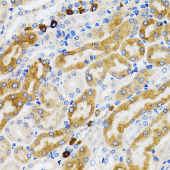
Immunohistochemistry analysis of paraffin-embedded Rat kidney using PGRMC1 Rabbit pAb (A5619) at dilution of 1:100 (40x lens). Microwave antigen retrieval performed with 0.01M PBS Buffer (pH 7.2) prior to IHC staining. |
|
|
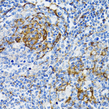
Immunohistochemistry analysis of paraffin-embedded Human tonsil using PGRMC1 Rabbit pAb (A5619) at dilution of 1:100 (40x lens). Microwave antigen retrieval performed with 0.01M PBS Buffer (pH 7.2) prior to IHC staining. |
|
|
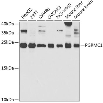
Western blot analysis of various lysates using PGRMC1 Rabbit pAb (A5619) at 1:1000 dilution. Secondary antibody: HRP-conjugated Goat anti-Rabbit IgG (H+L) (AS014) at 1:10000 dilution. Lysates/proteins: 25µg per lane. Blocking buffer: 3% nonfat dry milk in TBST. Detection: ECL Enhanced Kit (RM00021). Exposure time: 90s. |
|
|
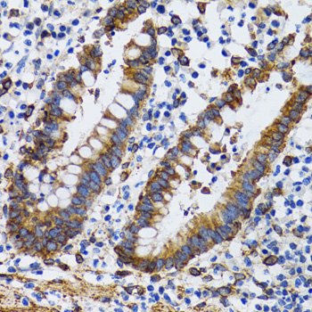
Immunohistochemistry analysis of paraffin-embedded Human vermiform appendix using PGRMC1 Rabbit pAb (A5619) at dilution of 1:100 (40x lens). Microwave antigen retrieval performed with 0.01M PBS Buffer (pH 7.2) prior to IHC staining. |
|
|
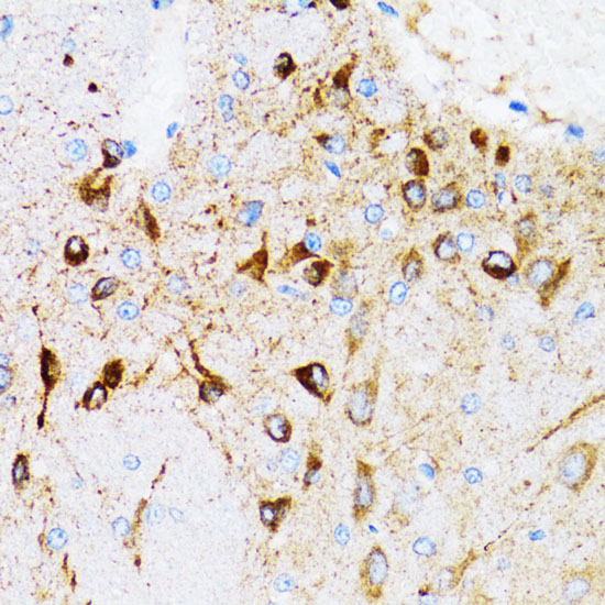
Immunohistochemistry analysis of paraffin-embedded Mouse brain using PGRMC1 Rabbit pAb (A5619) at dilution of 1:100 (40x lens). Microwave antigen retrieval performed with 0.01M PBS Buffer (pH 7.2) prior to IHC staining. |
|
|
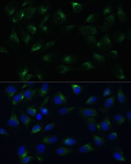
Immunofluorescence analysis of C6 cells using PGRMC1 Rabbit pAb (A5619) at dilution of 1:100 (40x lens). Secondary antibody: Cy3-conjugated Goat anti-Rabbit IgG (H+L) (AS007) at 1:500 dilution. Blue: DAPI for nuclear staining. |
|
|
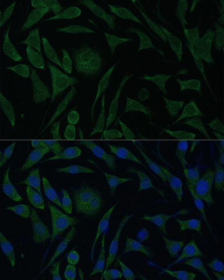
Immunofluorescence analysis of L929 cells using PGRMC1 Rabbit pAb (A5619) at dilution of 1:100 (40x lens). Secondary antibody: Cy3-conjugated Goat anti-Rabbit IgG (H+L) (AS007) at 1:500 dilution. Blue: DAPI for nuclear staining. |
|
|
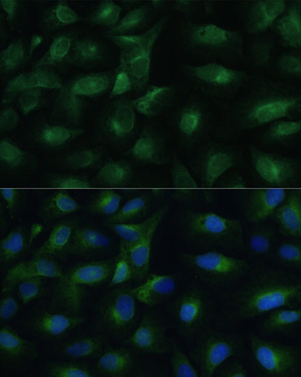
Immunofluorescence analysis of U-2 OS cells using PGRMC1 Rabbit pAb (A5619) at dilution of 1:100 (40x lens). Secondary antibody: Cy3-conjugated Goat anti-Rabbit IgG (H+L) (AS007) at 1:500 dilution. Blue: DAPI for nuclear staining. |
Produktgarantie und fachkundiger Support
