
ADAM15 Rabbit mAb, Unconjugated, Monoclonal
Artikelnummer:
ABB-A6813
- Bilder (9)

| Artikelname: | ADAM15 Rabbit mAb, Unconjugated, Monoclonal |
| Artikelnummer: | ABB-A6813 |
| Hersteller Artikelnummer: | A6813 |
| Alternativnummer: | ABB-A6813-100UL,ABB-A6813-20UL |
| Hersteller: | ABclonal |
| Wirt: | Rabbit |
| Kategorie: | Antikörper |
| Applikation: | ELISA, IF, IHC-P, WB |
| Spezies Reaktivität: | Human |
| Immunogen: | Synthetic peptide. This information is considered to be commercially sensitive. |
| Konjugation: | Unconjugated |
| Alternative Synonym: | MDC15, ADAM15 |
| The protein encoded by this gene is a member of the ADAM (a disintegrin and metalloproteinase) protein family. ADAM family members are type I transmembrane glycoproteins known to be involved in cell adhesion and proteolytic ectodomain processing of cytokines and adhesion molecules. This protein contains multiple functional domains including a zinc-binding metalloprotease domain, a disintegrin-like domain, as well as a EGF-like domain. Through its disintegrin-like domain, this protein specifically interacts with the integrin beta chain, beta 3. It also interacts with Src family protein-tyrosine kinases in a phosphorylation-dependent manner, suggesting that this protein may function in cell-cell adhesion as well as in cellular signaling. Multiple alternatively spliced transcript variants encoding distinct isoforms have been observed. |
| Application Verdünnung: | WB,1:1000 - 1:2000|IF-P,1:100 - 1:800|IHC-P,1:200 - 1:2000|ELISA,Recommended starting concentration is 1 µg/mL. Please optimize the concentration based on your specific assay requirements. |
| Anwendungsbeschreibung: | Cross-Reactivity: Human,Mouse,Rat. ResearchArea: Cancer,Invasion and Metastasis,Signal Transduction,Cell Biology Developmental Biology,Cell Adhesion,Cytoskeleton,Extracellular Matrix,Ubiquitin. Shipping: Ice Bag |
|
|
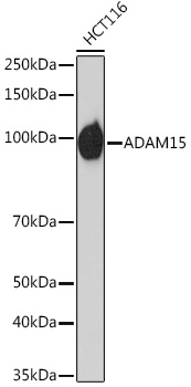
Western blot analysis of lysates from HCT116 cells, using ADAM15 Rabbit mAb (A6813) at 1:1000 dilution. Secondary antibody: HRP-conjugated Goat anti-Rabbit IgG (H+L) (AS014) at 1:10000 dilution. Lysates/proteins: 25µg per lane. Blocking buffer: 3% nonfat dry milk in TBST. Detection: ECL Basic Kit (RM00020). Exposure time: 60s. |
|
|
Immunohistochemistry analysis of paraffin-embedded Human colon carcinoma tissue using ADAM15 Rabbit mAb (A6813) at a dilution of 1:200 (40x lens). High pressure antigen retrieval performed with 0.01M Citrate buffer (pH 6.0) prior to IHC staining. |
|
|
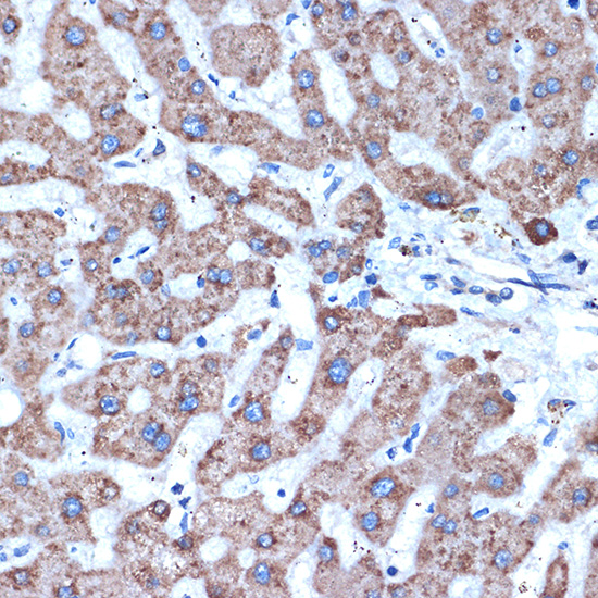
Immunohistochemistry analysis of paraffin-embedded Human liver tissue using ADAM15 Rabbit mAb (A6813) at a dilution of 1:200 (40x lens). High pressure antigen retrieval performed with 0.01M Citrate buffer (pH 6.0) prior to IHC staining. |
|
|
Immunohistochemistry analysis of paraffin-embedded Human small intestine tissue using ADAM15 Rabbit mAb (A6813) at a dilution of 1:200 (40x lens). High pressure antigen retrieval performed with 0.01M Citrate buffer (pH 6.0) prior to IHC staining. |
|
|
Immunohistochemistry analysis of paraffin-embedded Mouse intestin tissue using ADAM15 Rabbit mAb (A6813) at a dilution of 1:200 (40x lens). High pressure antigen retrieval performed with 0.01M Citrate buffer (pH 6.0) prior to IHC staining. |
|
|
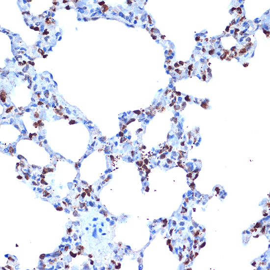
Immunohistochemistry analysis of paraffin-embedded Rat lung tissue using ADAM15 Rabbit mAb (A6813) at a dilution of 1:200 (40x lens). High pressure antigen retrieval performed with 0.01M Citrate buffer (pH 6.0) prior to IHC staining. |
|
|
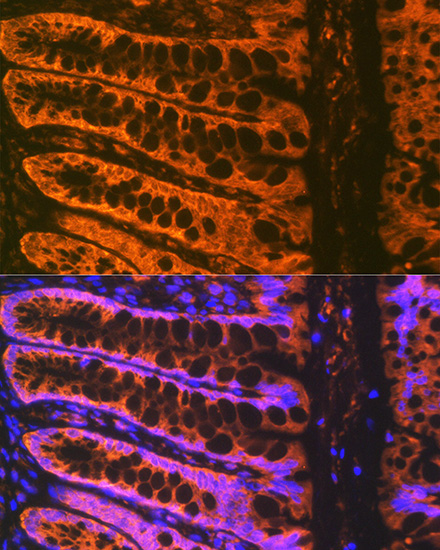
Immunofluorescence analysis of paraffin-embedded Rat rectum tissue using ADAM15 Rabbit mAb (A6813) at dilution of 1:100 (40x lens). Secondary antibody: Cy3-conjugated Goat anti-Rabbit IgG (H+L) (AS007) at 1:500 dilution. Blue: DAPI for nuclear staining. High pressure antigen retrieval performed with 0.01M Citrate Buffer (pH 6.0) prior to IF staining. |
|
|
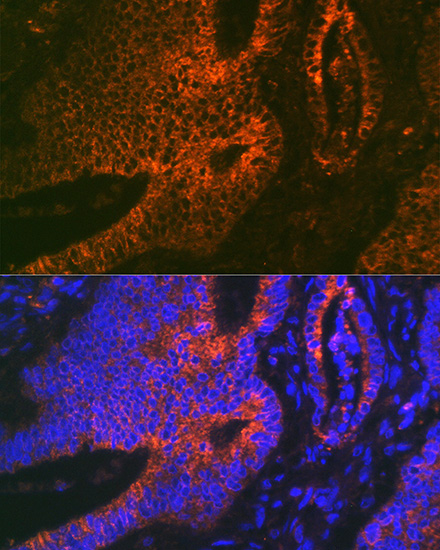
Confocal imaging of paraffin-embedded Human colon cancer tissue using ADAM15 Rabbit mAb (A6813, dilution 1:200) followed by a further incubation with Cy3 Goat Anti-Rabbit IgG (H+L) (AS007, dilution 1:500)(Red). DAPI was used for nuclear staining (Blue). Objective: 40x. High pressure antigen retrieval performed with 0.01M Citrate Buffer (pH 6.0) prior to IF staining. |
|
|
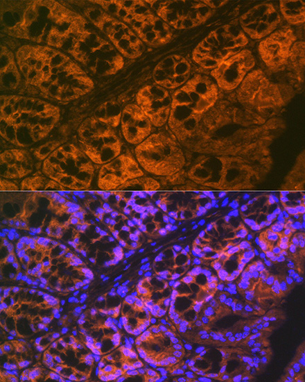
Confocal imaging of paraffin-embedded Mouse large intestine tissue using ADAM15 Rabbit mAb (A6813,dilution 1:200) followed by a further incubation with Cy3 Goat Anti-Rabbit IgG (H+L) (AS007,dilution 1:500)(Red).DAPI was used for nuclear staining (Blue). Objective: 40x. High pressure antigen retrieval performed with 0.01M Citrate Buffer (pH 6.0) prior to IF staining. |
Produktgarantie und fachkundiger Support
