
RPL26L1 Rabbit mAb, Unconjugated, Monoclonal
Artikelnummer:
ABB-A9419
- Bilder (8)


| Artikelname: | RPL26L1 Rabbit mAb, Unconjugated, Monoclonal |
| Artikelnummer: | ABB-A9419 |
| Hersteller Artikelnummer: | A9419 |
| Alternativnummer: | ABB-A9419-20UL,ABB-A9419-100UL |
| Hersteller: | ABclonal |
| Wirt: | Rabbit |
| Kategorie: | Antikörper |
| Applikation: | ELISA, IF, IHC-P, WB |
| Spezies Reaktivität: | Human |
| Immunogen: | Synthetic peptide. This information is considered to be commercially sensitive. |
| Konjugation: | Unconjugated |
| Alternative Synonym: | RPL26P1, RPL26L1 |
| This gene encodes a protein that shares high sequence similarity with ribosomal protein L26. Alternative splicing results in multiple transcript variants encoding the same protein. |
| Application Verdünnung: | WB,1:1000 - 1:6000|IF/ICC,1:50 - 1:200|IF-P,1:50 - 1:200|IHC-P,1:800 - 1:3200|ELISA,Recommended starting concentration is 1 µg/mL. Please optimize the concentration based on your specific assay requirements. |
| Anwendungsbeschreibung: | Cross-Reactivity: Human,Mouse,Rat. ResearchArea: Epigenetics Nuclear Signaling,Cell Biology Developmental Biology. Shipping: Ice Bag |
|
|
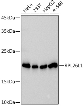
Western blot analysis of various lysates using (A9419) at 1:1000 dilution. Secondary antibody: HRP-conjugated Goat anti-Rabbit IgG (H+L) (AS014) at 1:10000 dilution. Lysates/proteins: 25µg per lane. Blocking buffer: 3% nonfat dry milk in TBST. Detection: ECL Basic Kit (RM00020). Exposure time: 3s. |
|
|
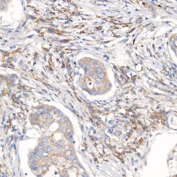
Immunohistochemistry analysis of paraffin-embedded Human colon carcinoma tissue using RPL26L1 Rabbit mAb (A9419) at a dilution of 1:1000 (40x lens). High pressure antigen retrieval performed with 0.01M Tris-EDTA Buffer (pH 9.0) prior to IHC staining. |
|
|
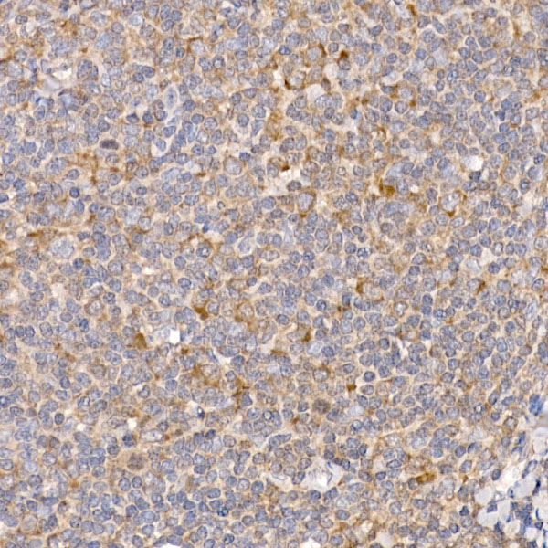
Immunohistochemistry analysis of paraffin-embedded Human tonsil tissue using RPL26L1 Rabbit mAb (A9419) at a dilution of 1:1000 (40x lens). High pressure antigen retrieval performed with 0.01M Tris-EDTA Buffer (pH 9.0) prior to IHC staining. |
|
|
Immunohistochemistry analysis of paraffin-embedded Mouse brain tissue using RPL26L1 Rabbit mAb (A9419) at a dilution of 1:1000 (40x lens). High pressure antigen retrieval performed with 0.01M Tris-EDTA Buffer (pH 9.0) prior to IHC staining. |
|
|
Immunohistochemistry analysis of paraffin-embedded Rat brain tissue using RPL26L1 Rabbit mAb (A9419) at a dilution of 1:1000 (40x lens). High pressure antigen retrieval performed with 0.01M Tris-EDTA Buffer (pH 9.0) prior to IHC staining. |
|
|
Confocal imaging of paraffin-embedded Mouse brain tissue using RPL26L1 Rabbit mAb (A9419, dilution 1:100) followed by a further incubation with Cy3 Goat Anti-Rabbit IgG (H+L) (AS007, dilution 1:500) (Red). DAPI was used for nuclear staining (Blue). Microwave antigen retrieval performed with 0.01M Citrate Buffer (pH 6.0) prior to IF staining. Objective: 40x. |
|
|
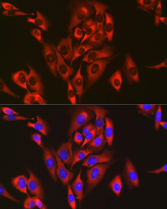
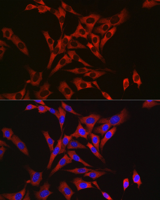
Confocal imaging of U-2 OS cells using RPL26L1 Rabbit mAb (A9419, dilution 1:50) followed by a further incubation with Cy3 Goat Anti-Rabbit IgG (H+L) (AS007, dilution 1:500) (Red). The cells were counterstained with alpha-Tubulin Mouse mAb (AC012, dilution 1:400) followed by incubation with ABflo 488-conjugated Goat Anti-Mouse IgG (H+L) Ab (AS076, dilution 1:500) (Green). DAPI was used for nuclear staining (Blue). Objective: 100x. |
|
|
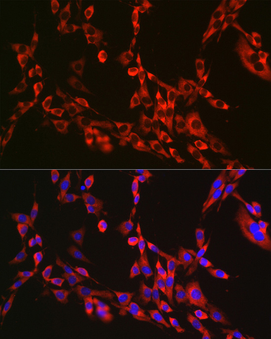
Confocal imaging of MCF7 cells using RPL26L1 Rabbit mAb (A9419, dilution 1:50) followed by a further incubation with Cy3 Goat Anti-Rabbit IgG (H+L) (AS007, dilution 1:500) (Red). The cells were counterstained with alpha-Tubulin Mouse mAb (AC012, dilution 1:400) followed by incubation with ABflo 488-conjugated Goat Anti-Mouse IgG (H+L) Ab (AS076, dilution 1:500) (Green). DAPI was used for nuclear staining (Blue). Objective: 100x. |
Produktgarantie und fachkundiger Support
