
Chromogranin A Rabbit mAb, Unconjugated, Monoclonal
Artikelnummer:
ABB-A9576
- Bilder (9)

| Artikelname: | Chromogranin A Rabbit mAb, Unconjugated, Monoclonal |
| Artikelnummer: | ABB-A9576 |
| Hersteller Artikelnummer: | A9576 |
| Alternativnummer: | ABB-A9576-20UL,ABB-A9576-100UL,ABB-A9576-1000UL,ABB-A9576-500UL |
| Hersteller: | ABclonal |
| Wirt: | Rabbit |
| Kategorie: | Antikörper |
| Applikation: | ELISA, IF, IHC-P |
| Spezies Reaktivität: | Human |
| Immunogen: | Synthetic peptide. This information is considered to be commercially sensitive. |
| Konjugation: | Unconjugated |
| Alternative Synonym: | CGA, PHE5, PHES, Chromogranin A |
| The protein encoded by this gene is a member of the chromogranin/secretogranin family of neuroendocrine secretory proteins. It is found in secretory vesicles of neurons and endocrine cells. This gene product is a precursor to three biologically active peptides, vasostatin, pancreastatin, and parastatin. These peptides act as autocrine or paracrine negative modulators of the neuroendocrine system. Two other peptides, catestatin and chromofungin, have antimicrobial activity and antifungal activity, respectively. Two transcript variants encoding different isoforms have been found for this gene. |
| Application Verdünnung: | IF-P,1:50 - 1:200|IHC-P,1:1000 - 1:5000|ELISA,Recommended starting concentration is 1 µg/mL. Please optimize the concentration based on your specific assay requirements. |
| Anwendungsbeschreibung: | Cross-Reactivity: Human,Mouse,Rat. ResearchArea: Cancer,Tumor biomarkers,Signal Transduction,Cell Biology Developmental Biology,Growth factors,Neuroscience, Cell Type Marker,Neuron marker. Shipping: Ice Bag |
|
|
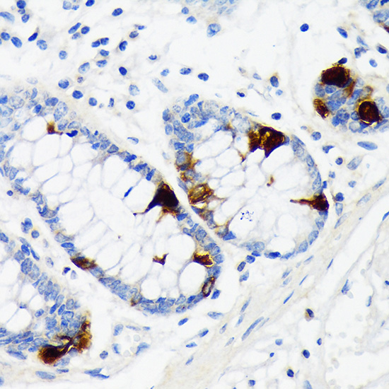
Immunohistochemistry analysis of paraffin-embedded Human colon tissue using Chromogranin A Rabbit mAb (A9576) at a dilution of 1:4000 (40x lens). High pressure antigen retrieval performed with 0.01M Citrate Buffer (pH 6.0) prior to IHC staining. |
|
|
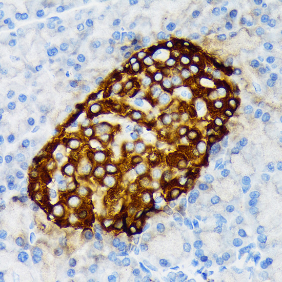
Immunohistochemistry analysis of paraffin-embedded Human pancreas tissue using Chromogranin A Rabbit mAb (A9576) at a dilution of 1:4000 (40x lens). High pressure antigen retrieval performed with 0.01M Citrate Buffer (pH 6.0) prior to IHC staining. |
|
|
Immunohistochemistry analysis of paraffin-embedded Rat colon tissue using Chromogranin A Rabbit mAb (A9576) at a dilution of 1:4000 (40x lens). High pressure antigen retrieval performed with 0.01M Citrate Buffer (pH 6.0) prior to IHC staining. |
|
|
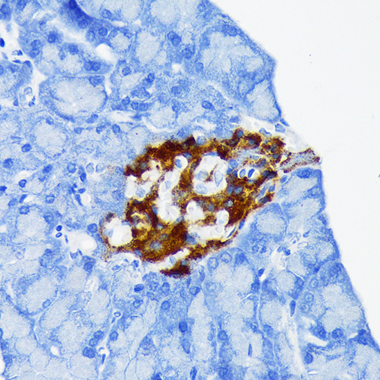
Immunohistochemistry analysis of paraffin-embedded Human pheochromocytoma tissue using Chromogranin A Rabbit mAb (A9576) at a dilution of 1:4000 (40x lens). High pressure antigen retrieval performed with 0.01M Citrate Buffer (pH 6.0) prior to IHC staining. |
|
|
Immunohistochemistry analysis of paraffin-embedded Mouse brain tissue using Chromogranin A Rabbit mAb (A9576) at a dilution of 1:4000 (40x lens). High pressure antigen retrieval performed with 0.01M Citrate Buffer (pH 6.0) prior to IHC staining. |
|
|
Immunohistochemistry analysis of paraffin-embedded Mouse colon tissue using Chromogranin A Rabbit mAb (A9576) at a dilution of 1:4000 (40x lens). High pressure antigen retrieval performed with 0.01M Citrate Buffer (pH 6.0) prior to IHC staining. |
|
|
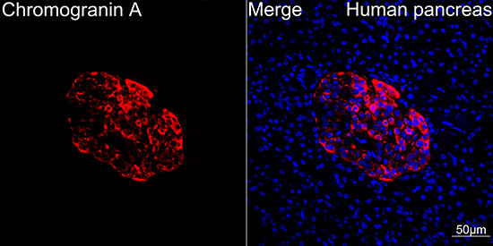
Confocal imaging of paraffin-embedded Human pancreas tissue using Chromogranin A Rabbit mAb (A9576, dilution 1:100) followed by a further incubation with Cy3 Goat Anti-Rabbit IgG (H+L) (AS007, dilution 1:500) (Red). DAPI was used for nuclear staining (Blue). High pressure antigen retrieval performed with 0.01M Citrate Buffer (pH 6.0) prior to IF staining. Objective: 40x. |
|
|
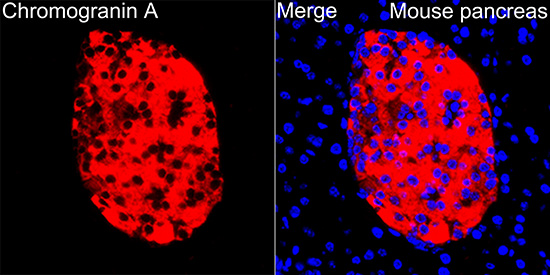
Confocal imaging of paraffin-embedded Mouse pancreas tissue using Chromogranin A Rabbit mAb (A9576, dilution 1:100) followed by a further incubation with Cy3 Goat Anti-Rabbit IgG (H+L) (AS007, dilution 1:500) (Red). DAPI was used for nuclear staining (Blue). High pressure antigen retrieval performed with 0.01M Citrate Buffer (pH 6.0) prior to IF staining. Objective: 40x. |
|
|
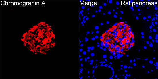
Confocal imaging of paraffin-embedded Rat pancreas tissue using Chromogranin A Rabbit mAb (A9576, dilution 1:100) followed by a further incubation with Cy3 Goat Anti-Rabbit IgG (H+L) (AS007, dilution 1:500) (Red). DAPI was used for nuclear staining (Blue). High pressure antigen retrieval performed with 0.01M Citrate Buffer (pH 6.0) prior to IF staining. Objective: 40x. |
Produktgarantie und fachkundiger Support
