
PSMD14 Rabbit mAb, Unconjugated, Monoclonal
Artikelnummer:
ABB-A9608
- Bilder (9)

| Artikelname: | PSMD14 Rabbit mAb, Unconjugated, Monoclonal |
| Artikelnummer: | ABB-A9608 |
| Hersteller Artikelnummer: | A9608 |
| Alternativnummer: | ABB-A9608-20UL,ABB-A9608-100UL |
| Hersteller: | ABclonal |
| Wirt: | Rabbit |
| Kategorie: | Antikörper |
| Applikation: | ELISA, IF, IHC-P, WB |
| Spezies Reaktivität: | Human |
| Immunogen: | Recombinant protein (or fragment).This information is considered to be commercially sensitive. |
| Konjugation: | Unconjugated |
| Alternative Synonym: | PAD1, POH1, RPN11, PSMD14 |
| This gene encodes a component of the 26S proteasome. The 26S proteasome is a large multiprotein complex that catalyzes the degradation of ubiquitinated intracellular proteins. The encoded protein is a component of the 19S regulatory cap complex of the 26S proteasome and mediates substrate deubiquitination. A pseudogene of this gene is also located on the long arm of chromosome 2. |
| Application Verdünnung: | WB,1:1000 - 1:6000|IHC-P,1:200 - 1:2000|IF/ICC,1:50 - 1:200|ELISA,Recommended starting concentration is 1 µg/mL. Please optimize the concentration based on your specific assay requirements. |
| Anwendungsbeschreibung: | Cross-Reactivity: Human,Mouse,Rat. ResearchArea: Epigenetics Nuclear Signaling,Cell Biology Developmental Biology,Ubiquitin. Shipping: Ice Bag |
|
|
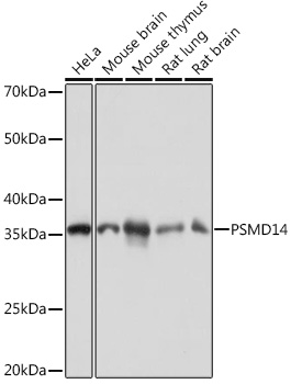
Western blot analysis of various lysates using PSMD14 Rabbit mAb (A9608) at 1:1000 dilution incubated overnight at 4°C. Secondary antibody: HRP-conjugated Goat anti-Rabbit IgG (H+L) (AS014) at 1:10000 dilution. Lysates/proteins: 25 µg per lane. Blocking buffer: 3% nonfat dry milk in TBST. Detection: ECL Basic Kit (RM00020). Exposure time: 10s. |
|
|
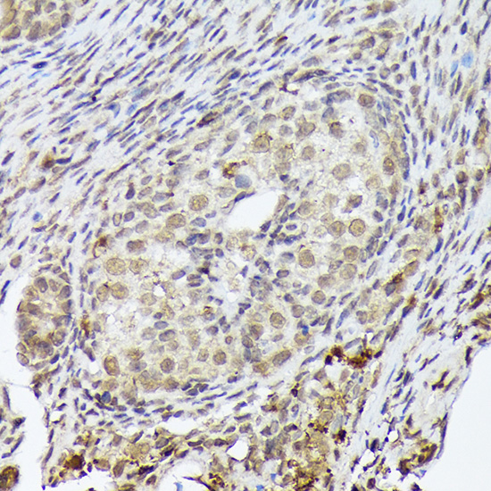
Immunohistochemistry analysis of paraffin-embedded Human cervix cancer tissue using PSMD14 Rabbit mAb (A9608) at a dilution of 1:200 (40x lens). High pressure antigen retrieval performed with 0.01M Citrate buffer (pH 6.0) prior to IHC staining. |
|
|
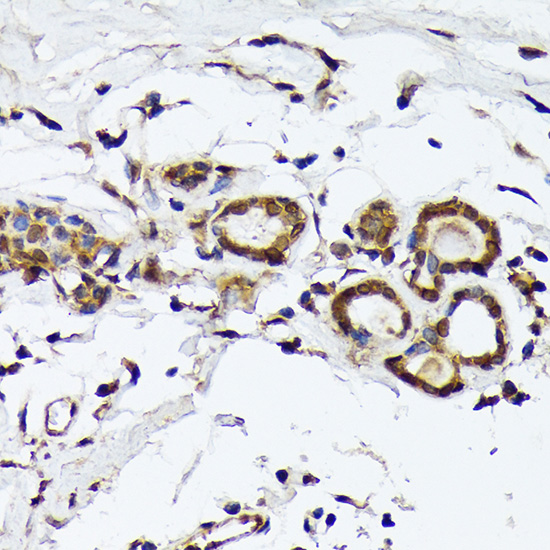
Immunohistochemistry analysis of paraffin-embedded Human colon carcinoma tissue using PSMD14 Rabbit mAb (A9608) at a dilution of 1:200 (40x lens). High pressure antigen retrieval performed with 0.01M Citrate buffer (pH 6.0) prior to IHC staining. |
|
|
Immunohistochemistry analysis of paraffin-embedded Human colon tissue using PSMD14 Rabbit mAb (A9608) at a dilution of 1:200 (40x lens). High pressure antigen retrieval performed with 0.01M Citrate buffer (pH 6.0) prior to IHC staining. |
|
|
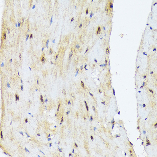
Immunohistochemistry analysis of paraffin-embedded Mouse brain tissue using PSMD14 Rabbit mAb (A9608) at a dilution of 1:200 (40x lens). High pressure antigen retrieval performed with 0.01M Citrate buffer (pH 6.0) prior to IHC staining. |
|
|
Immunohistochemistry analysis of paraffin-embedded Mouse intestin tissue using PSMD14 Rabbit mAb (A9608) at a dilution of 1:200 (40x lens). High pressure antigen retrieval performed with 0.01M Citrate buffer (pH 6.0) prior to IHC staining. |
|
|
Immunohistochemistry analysis of paraffin-embedded Mouse kidney tissue using PSMD14 Rabbit mAb (A9608) at a dilution of 1:200 (40x lens). High pressure antigen retrieval performed with 0.01M Citrate buffer (pH 6.0) prior to IHC staining. |
|
|
Immunohistochemistry analysis of paraffin-embedded Rat colon tissue using PSMD14 Rabbit mAb (A9608) at a dilution of 1:200 (40x lens). High pressure antigen retrieval performed with 0.01M Citrate buffer (pH 6.0) prior to IHC staining. |
|
|
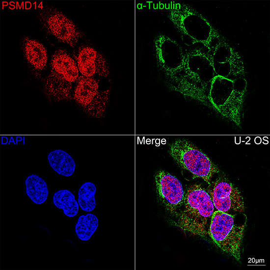
Confocal imaging of U-2OS cells using PSMD14 Rabbit mAb (A9608, dilution 1:100) (Red). The cells were counterstained with alpha-Tubulin Mouse mAb (AC012, dilution 1:400) (Green). DAPI was used for nuclear staining (blue). Objective: 40x. |
Produktgarantie und fachkundiger Support
