
alpha-Tubulin Rabbit mAb, Unconjugated
Artikelnummer:
ABB-AC049
- Bilder (8)


| Artikelname: | alpha-Tubulin Rabbit mAb, Unconjugated |
| Artikelnummer: | ABB-AC049 |
| Hersteller Artikelnummer: | AC049 |
| Alternativnummer: | ABB-AC049-100UL,ABB-AC049-200UL,ABB-AC049-1000UL,ABB-AC049-50UL,ABB-AC049-500UL |
| Hersteller: | ABclonal |
| Wirt: | Rabbit |
| Kategorie: | Antikörper |
| Applikation: | ELISA, IF, IHC-P, WB |
| Spezies Reaktivität: | Human |
| Immunogen: | Recombinant protein (or fragment).This information is considered to be commercially sensitive. |
| Konjugation: | Unconjugated |
| Alternative Synonym: | K-ALPHA-1, alpha-Tubulin |
| Enables double-stranded RNA binding activity and ubiquitin protein ligase binding activity. Predicted to be involved in microtubule cytoskeleton organization and mitotic cell cycle. Predicted to act upstream of or within cellular response to interleukin-4. Located in microtubule. |
| Application Verdünnung: | WB,1:10000 - 1:50000|IF/ICC,1:500 - 1:2000|IHC-P,1:200 - 1:1000|ELISA,Recommended starting concentration is 1 µg/mL. Please optimize the concentration based on your specific assay requirements. |
| Anwendungsbeschreibung: | Cross-Reactivity: Human,Mouse,Rat. ResearchArea: Signal Transduction,Cell Biology Developmental Biology,Cell Cycle,Centrosome,Cytoskeleton,Microtubules. Shipping: Ice Bag |
|
|
Immunohistochemistry analysis of paraffin-embedded Human cervix cancer tissue using alpha-Tubulin Rabbit mAb (AC049) at a dilution of 1:200 (40x lens). High pressure antigen retrieval performed with 0.01M Citrate buffer (pH 6.0) prior to IHC staining. |
|
|
Immunohistochemistry analysis of paraffin-embedded Human colon carcinoma tissue using alpha-Tubulin Rabbit mAb (AC049) at a dilution of 1:200 (40x lens). High pressure antigen retrieval performed with 0.01M Citrate buffer (pH 6.0) prior to IHC staining. |
|
|
Western blot analysis of various lysates using alpha-Tubulin Rabbit mAb (AC049) at 1:50000 dilution incubated overnight at 4°C. Secondary antibody: HRP-conjugated Goat anti-Rabbit IgG (H+L) (AS014) at 1:10000 dilution. Lysates/proteins: 25 µg per lane. Blocking buffer: 3% nonfat dry milk in TBST. Detection: ECL Basic Kit (RM00020). Exposure time: 30s. |
|
|
Immunohistochemistry analysis of paraffin-embedded Human tonsil tissue using alpha-Tubulin Rabbit mAb (AC049) at a dilution of 1:200 (40x lens). High pressure antigen retrieval performed with 0.01M Citrate buffer (pH 6.0) prior to IHC staining. |
|
|
Immunohistochemistry analysis of paraffin-embedded Mouse kidney tissue using alpha-Tubulin Rabbit mAb (AC049) at a dilution of 1:200 (40x lens). High pressure antigen retrieval performed with 0.01M Citrate buffer (pH 6.0) prior to IHC staining. |
|
|
Immunohistochemistry analysis of paraffin-embedded Mouse testis tissue using alpha-Tubulin Rabbit mAb (AC049) at a dilution of 1:200 (40x lens). High pressure antigen retrieval performed with 0.01M Citrate buffer (pH 6.0) prior to IHC staining. |
|
|
Immunohistochemistry analysis of paraffin-embedded Rat colon tissue using alpha-Tubulin Rabbit mAb (AC049) at a dilution of 1:200 (40x lens). High pressure antigen retrieval performed with 0.01M Citrate buffer (pH 6.0) prior to IHC staining. |
|
|
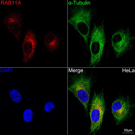
Confocal imaging of HeLa cells using alpha-Tubulin Rabbit mAb (AC049, dilution 1:1000) followed by a further incubation with Cy3-conjugated Goat Anti-Rabbit IgG (H+L) (AS007, dilution 1:500) (Red). The cells were counterstained with alpha-Tubulin Mouse mAb (AC012, dilution 1:400) followed by incubation with ABflo 488-conjugated Goat Anti-Mouse IgG (H+L) (AS076, dilution 1:500) (Green). DAPI was used for nuclear staining (Blue). Objective: 100x. |
Produktgarantie und fachkundiger Support
