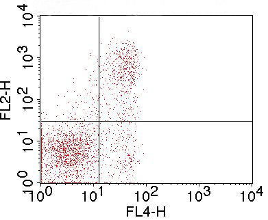
Figure-1: Cell surface flowcytometric staining of mFR4 (Clone: TH6) in mouse Thymocytes. Mouse Thymocytes were first stained with Anti-mCD3 APC conjugated, mCD3 APC positive cells were gated and further analyzed for mFR4 using 0.5 µg/10 6. Goat anti-Rat PE secondary antibody (ABEOMICS) was used.
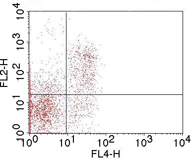
Figure-2: Cell surface flowcytometric staining of mFR4 (Clone: TH6) in mouse Splenocytes. Mouse Splenocytes were first stained with Anti-mCD3 APC conjugated, mCD3 APC positive cells were gated and further analyzed for mFR4 using 0.5 µg/10 6. Goat anti-Rat PE secondary antibody (ABEOMICS) was used.
* Mehrwertsteuer und Versandkosten nicht enthalten. Irrtümer und Preisänderungen vorbehalten