Western blot analysis: 4-6 µg/ml, Flowcytometric analysis: 2-4 µg/10 6 Cells
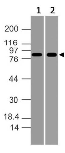
Figure-1: Western blot analysis of MLH1. Anti- MLH1 antibody (Clone: ABM17C1) was used at 4 µg/ml on (1) MCF-7 and (2) Raji lysates.
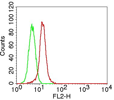
Figure-2: Intracellular flow cytometric analysis of MLH1 in A431 cells using 2 µg/10 6 cells of Anti-MLH1 (Clone: ABM17C1). Green represent isotype control and red represent Anti MLH1 antibody. PE conjugate Goat anti mouse IgG was used as secondary control
* Mehrwertsteuer und Versandkosten nicht enthalten. Irrtümer und Preisänderungen vorbehalten