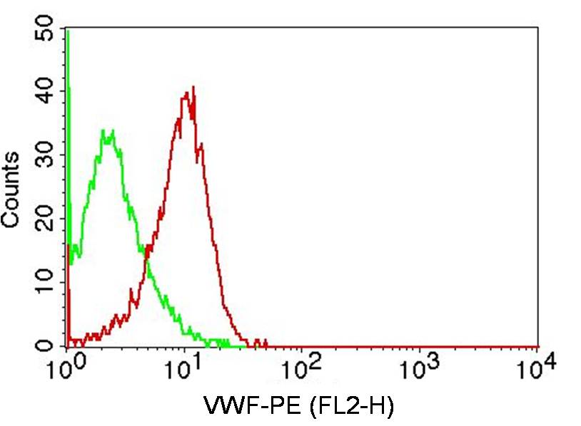
Fig-1: Intracellular flow analysis of VWF antibody on HepG2 cells using 0.5 µg/10 6 cells of antibody (Clone: ABM53D2). Green represents isotype control, red represents anti-VWF antibody. Goat anti-mouse PE conjugate was used as secondary antibody. (Cells were fixed with 4% paraformaldehyde for 10 min and washed with PBS by centrifuging at 1100 for 5 min followed by permeabilization for 20 min and washed again as mentioned above. Then cell were incubated with primary antibody for 45 min. and
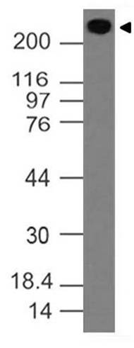
Fig-2: Western blot analysis of VWF. Anti-VWF antibody (Clone : ABM53D2) was used at 2 µg/ml on h Lungs lysate.
* Mehrwertsteuer und Versandkosten nicht enthalten. Irrtümer und Preisänderungen vorbehalten