E.coli-derived human BCLAF1 recombinant protein (Position: H293-E901). Human BCLAF1 shares 94.4% amino acid (aa) sequence identity with mouse BCLAF1.
Alternative Synonym:
BCLAF1, BTF, KIAA0164, Bcl-2-associated transcription factor 1, Btf, BCLAF1 and THRAP3 family member 1
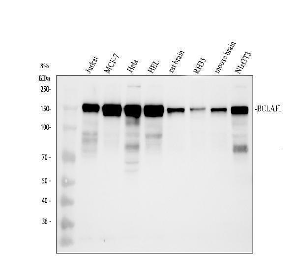
Boster Bio Anti-BCLAF1 Antibody Picoband catalog A04100-3. Tested in WB, IHC, ICC, IF, IP, Flow Cytometry, ELISA applications. This antibody reacts with Human, Mouse, Rat. The brand Picoband indicates this is a premium antibody that guarantees superior quality, high affinity, and strong signals with minimal background in Western blot applications. Only our best-performing antibodies are designated as Picoband, ensuring unmatched performance.
Klonalität:
Polyclonal
Konzentration:
Adding 0.2 ml of distilled water will yield a concentration of 500 µg/ml.
Western blot, 0.25-0.5 µg/ml, Human, Mouse, Rat Immunohistochemistry (Paraffin-embedded Section), 2-5 µg/ml, Human, Mouse, Rat Immunocytochemistry/Immunofluorescence, 5 µg/ml, Human Immunoprecipitation, 0.5-2 µg/ml, Human Flow Cytometry (Fixed), 1-3 µg/1x
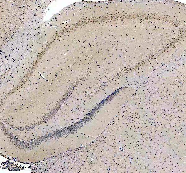
IHC analysis of BCLAF1 using anti-BCLAF1 antibody (A04100-3). BCLAF1 was detected in a paraffin-embedded section of rat brain tissue. Heat mediated antigen retrieval was performed in EDTA buffer (pH 8.0, epitope retrieval solution). The tissue section was blocked with 10% goat serum. The tissue section was then incubated with 2 µg/ml rabbit anti-BCLAF1 Antibody (A04100-3) overnight at 4C. Peroxidase Conjugated Goat Anti-rabbit IgG was used as secondary antibody and incubated for 30 minutes at 37C. The tissue section was developed using HRP Conjugated Rabbit IgG Super Vision Assay Kit (Catalog SV0002) with DAB as the chromogen.
IHC analysis of BCLAF1 using anti-BCLAF1 antibody (A04100-3). BCLAF1 was detected in a paraffin-embedded section of rat brain tissue. Heat mediated antigen retrieval was performed in EDTA buffer (pH 8.0, epitope retrieval solution). The tissue section was blocked with 10% goat serum. The tissue section was then incubated with 2 µg/ml rabbit anti-BCLAF1 Antibody (A04100-3) overnight at 4C. Peroxidase Conjugated Goat Anti-rabbit IgG was used as secondary antibody and incubated for 30 minutes at 37C. The tissue section was developed using HRP Conjugated Rabbit IgG Super Vision Assay Kit (Catalog SV0002) with DAB as the chromogen.
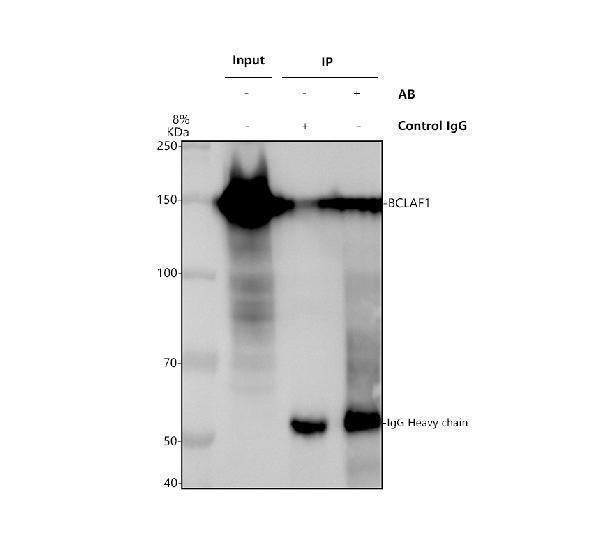
Immunoprecipitating BCLAF1 in Hela whole cell lysate.Western blot analysis of BCLAF1 using anti-BCLAF1 antibody (A04100-3).Lane 1: Hela whole cell lysates (30ug),Lane 2: Rabbit control IgG instead of anti-BCLAF1 antibody in Hela whole cell lysate,Lane 3: anti-BCLAF1 antibody (2µg) + Hela whole cell lysate (500µg).After electrophoresis, proteins were transferred to a membrane. Then the membrane was incubated with rabbit anti-BCLAF1 antigen affinity purified polyclonal antibody (A04100-3) at a dilution of 0.5 µg/mL and probed with a goat anti-rabbit IgG-HRP secondary antibody (Catalog BA1054). The signal is developed using ECL Plus Western Blotting Su
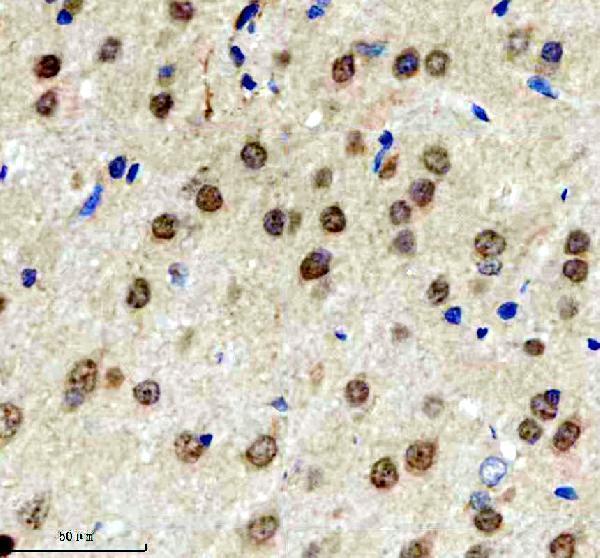
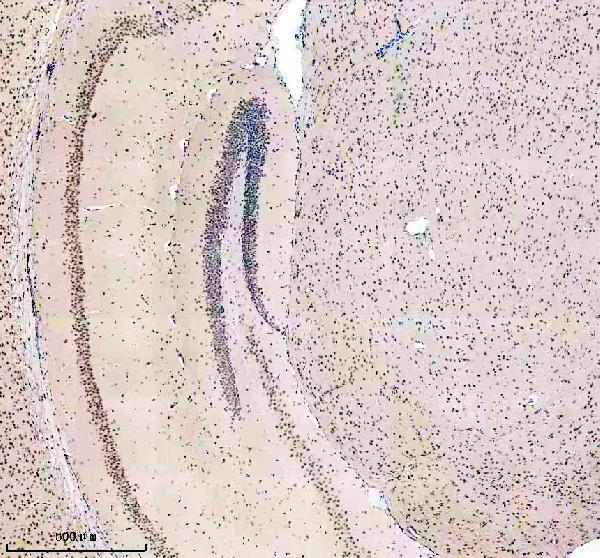
IHC analysis of BCLAF1 using anti-BCLAF1 antibody (A04100-3). BCLAF1 was detected in a paraffin-embedded section of mouse brain tissue. Heat mediated antigen retrieval was performed in EDTA buffer (pH 8.0, epitope retrieval solution). The tissue section was blocked with 10% goat serum. The tissue section was then incubated with 2 µg/ml rabbit anti-BCLAF1 Antibody (A04100-3) overnight at 4C. Peroxidase Conjugated Goat Anti-rabbit IgG was used as secondary antibody and incubated for 30 minutes at 37C. The tissue section was developed using HRP Conjugated Rabbit IgG Super Vision Assay Kit (Catalog SV0002) with DAB as the chromogen.
IF analysis of BCLAF1 using anti-BCLAF1 antibody (A04100-3) and anti-Beta Tubulin antibody (M01857-3). BCLAF1 was detected in an immunocytochemical section of Hela cells. Enzyme antigen retrieval was performed using IHC enzyme antigen retrieval reagent (AR0022) for 15 mins. The cells were blocked with 10% goat serum. And then incubated with 5 µg/mL rabbit anti-BCLAF1 Antibody (A04100-3) and mouse anti-Beta Tubulin antibody (M01857-3) overnight at 4C. DyLight488 Conjugated Goat Anti-Rabbit IgG (BA1127) and Cy3 Conjugated Goat Anti-Mouse IgG (BA1031) were used as secondary antibody at 1:500 dilution and incubated for 30 minutes at 37C. Visualize using a fluorescence microscope and filter sets appropriate for the label used.
IHC analysis of BCLAF1 using anti-BCLAF1 antibody (A04100-3). BCLAF1 was detected in a paraffin-embedded section of mouse brain tissue. Heat mediated antigen retrieval was performed in EDTA buffer (pH 8.0, epitope retrieval solution). The tissue section was blocked with 10% goat serum. The tissue section was then incubated with 2 µg/ml rabbit anti-BCLAF1 Antibody (A04100-3) overnight at 4C. Peroxidase Conjugated Goat Anti-rabbit IgG was used as secondary antibody and incubated for 30 minutes at 37C. The tissue section was developed using HRP Conjugated Rabbit IgG Super Vision Assay Kit (Catalog SV0002) with DAB as the chromogen.
* Mehrwertsteuer und Versandkosten nicht enthalten. Irrtümer und Preisänderungen vorbehalten