
ATG3 Antibody, Unconjugated, Rabbit, Polyclonal
Artikelnummer:
CSB-PA002288HA01HU
- Bilder (4)
| Artikelname: | ATG3 Antibody, Unconjugated, Rabbit, Polyclonal |
| Artikelnummer: | CSB-PA002288HA01HU |
| Hersteller Artikelnummer: | CSB-PA002288HA01HU |
| Alternativnummer: | CSB-PA002288HA01HU-100UG, CSB-PA002288HA01HU-50UG |
| Hersteller: | Cusabio |
| Wirt: | Rabbit |
| Kategorie: | Antikörper |
| Applikation: | ELISA, IF, IHC, WB |
| Spezies Reaktivität: | Human |
| Konjugation: | Unconjugated |
| Alternative Synonym: | 2610016C12Rik antibody, Apg 3 antibody, APG3 antibody, APG3 autophagy 3 like antibody, APG3 like antibody, APG3, S. cerevisiae, homolog of antibody, APG3-like antibody, APG3L antibody, Apg3p antibody, ATG 3 antibody, ATG3 antibody, ATG3 autophagy related 3 homolog antibody, ATG3 autophagy related 3 homolog (S. cerevisiae) antibody, ATG3_HUMAN antibody, Autophagy 3, S. cerevisiae, homolog of antibody, Autophagy Apg3p/Aut1p like antibody, autophagy related 3 antibody, Autophagy related protein 3 antibody, Autophagy-related protein 3 antibody, DKFZp564M1178 antibody, FLJ22125 antibody, hApg3 antibody, MGC15201 antibody, OTTHUMP00000214547 antibody, OTTHUMP00000214548 antibody, PC3 96 antibody, Protein PC3-96 antibody, Ubiquitin-like-conjugating enzyme ATG3 antibody |
| Klonalität: | Polyclonal |
| UniProt: | Q9NT62 |
| Puffer: | Preservative: 0.03% Proclin 300<br />Constituents: 50% Glycerol, 0.01M PBS, PH 7.4 |
| Reinheit: | >95%, Protein G purified |
| Formulierung: | Liquid |
| Target-Kategorie: | ATG3 |
| Application Verdünnung: | Recommended dilution: WB:1:500-1:5000, IHC:1:1000-1:2000, IF:1:200-1:500 |
|
|
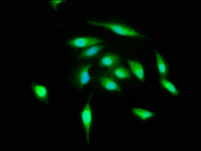
Immunofluorescence staining of Hela cells with CSB-PA002288HA01HU at 1:400, counter-stained with DAPI. The cells were fixed in 4% formaldehyde, permeabilized using 0.2% Triton X-100 and blocked in 10% normal Goat Serum. The cells were then incubated with the antibody overnight at 4°,C. The secondary antibody was Alexa Fluor 488-congugated AffiniPure Goat Anti-Rabbit IgG(H+L). |
|
|
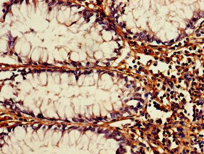
IHC image of CSB-PA002288HA01HU diluted at 1:1200 and staining in paraffin-embedded human colon cancer performed on a Leica BondTM system. After dewaxing and hydration, antigen retrieval was mediated by high pressure in a citrate buffer (pH 6.0). Section was blocked with 10% normal goat serum 30min at RT. Then primary antibody (1% BSA) was incubated at 4°,C overnight. The primary is detected by a biotinylated secondary antibody and visualized using an HRP conjugated SP system. |
|
|
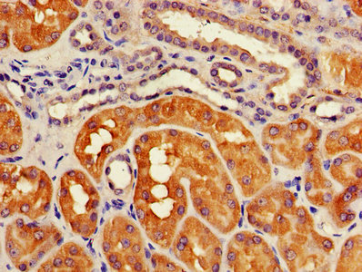
IHC image of CSB-PA002288HA01HU diluted at 1:1200 and staining in paraffin-embedded human kidney tissue performed on a Leica BondTM system. After dewaxing and hydration, antigen retrieval was mediated by high pressure in a citrate buffer (pH 6.0). Section was blocked with 10% normal goat serum 30min at RT. Then primary antibody (1% BSA) was incubated at 4°,C overnight. The primary is detected by a biotinylated secondary antibody and visualized using an HRP conjugated SP system. |
|
|
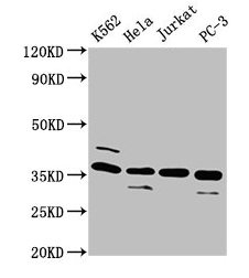
Western Blot Positive WB detected in: K562 whole cell lysate, Hela whole cell lysate, Jurkat whole cell lysate, PC-3 whole cell lysate All lanes: ATG3 antibody at 4µg/ml Secondary Goat polyclonal to rabbit IgG at 1/50000 dilution Predicted band size: 36 kDa Observed band size: 36 kDa |
Produktgarantie und fachkundiger Support
