
CBL Rabbit pAb, Unconjugated, Polyclonal
Catalog Number:
ABB-A0732
- Images (8)


| Article Name: | CBL Rabbit pAb, Unconjugated, Polyclonal |
| Biozol Catalog Number: | ABB-A0732 |
| Supplier Catalog Number: | A0732 |
| Alternative Catalog Number: | ABB-A0732-100UL,ABB-A0732-20UL,ABB-A0732-1000UL,ABB-A0732-500UL |
| Manufacturer: | ABclonal |
| Host: | Rabbit |
| Category: | Antikörper |
| Application: | ELISA, IF, IHC-P, IP, WB |
| Species Reactivity: | Human |
| Immunogen: | Recombinant protein (or fragment).This information is considered to be commercially sensitive. |
| Conjugation: | Unconjugated |
| Alternative Names: | CBL2, NSLL, C-CBL, RNF55, FRA11B, CBL |
| This gene is a proto-oncogene that encodes a RING finger E3 ubiquitin ligase. The encoded protein is one of the enzymes required for targeting substrates for degradation by the proteasome. This protein mediates the transfer of ubiquitin from ubiquitin conjugating enzymes (E2) to specific substrates. This protein also contains an N-terminal phosphotyrosine binding domain that allows it to interact with numerous tyrosine-phosphorylated substrates and target them for proteasome degradation. As such it functions as a negative regulator of many signal transduction pathways. This gene has been found to be mutated or translocated in many cancers including acute myeloid leukaemia, and expansion of CGG repeats in the 5 UTR has been associated with Jacobsen syndrome. Mutations in this gene are also the cause of Noonan syndrome-like disorder. |
| Application Dilute: | WB,1:1000 - 1:5000|IHC-P,1:50 - 1:100|IF/ICC,1:100 - 1:500|IP,0.5µg-4µg antibody for 400µg-600µg extracts of whole cells|ELISA,Recommended starting concentration is 1 µg/mL. Please optimize the concentration based on your specific assay requirements. |
| Application Notes: | Cross-Reactivity: Human,Mouse,Rat. ResearchArea: Epigenetics Nuclear Signaling,Nuclear Receptor Signaling,Nuclear hormone receptors,Protein phosphorylation,Cancer,Signal Transduction,ErbB-HER Signaling Pathway,Cell Biology Developmental Biology,Ubiquitin,Ubiquitin-Proteasome Signaling Pathway,Endocrine Metabolism,Insulin Receptor Signaling Pathway,Immunology Inflammation,B Cell Receptor Signaling Pathway,T Cell Receptor Signaling Pathway. Shipping: Ice Bag |
|
|
Western blot analysis of lysates from Mouse thymus, using CBL Rabbit pAb (A0732) at 1:3000 dilution. Secondary antibody: HRP-conjugated Goat anti-Rabbit IgG (H+L) (AS014) at 1:10000 dilution. Lysates/proteins: 25µg per lane. Blocking buffer: 3% nonfat dry milk in TBST. Detection: ECL Basic Kit (RM00020). Exposure time: 1s. |
|
|
Immunohistochemistry analysis of paraffin-embedded Human prostate using CBL Rabbit pAb (A0732) at dilution of 1:200 (40x lens). Microwave antigen retrieval performed with 0.01M PBS Buffer (pH 7.2) prior to IHC staining. |
|
|
Western blot analysis of lysates from 293T cells, using CBL Rabbit pAb (A0732) at 1:3000 dilution. Secondary antibody: HRP-conjugated Goat anti-Rabbit IgG (H+L) (AS014) at 1:10000 dilution. Lysates/proteins: 25µg per lane. Blocking buffer: 3% nonfat dry milk in TBST. Detection: ECL Basic Kit (RM00020). Exposure time: 30s. |
|
|
Immunohistochemistry analysis of paraffin-embedded Mouse lung using CBL Rabbit pAb (A0732) at dilution of 1:200 (40x lens). Microwave antigen retrieval performed with 0.01M PBS Buffer (pH 7.2) prior to IHC staining. |
|
|
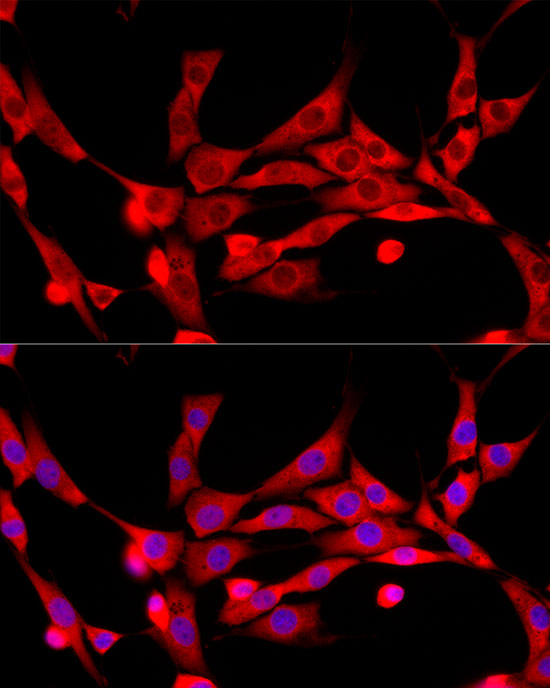
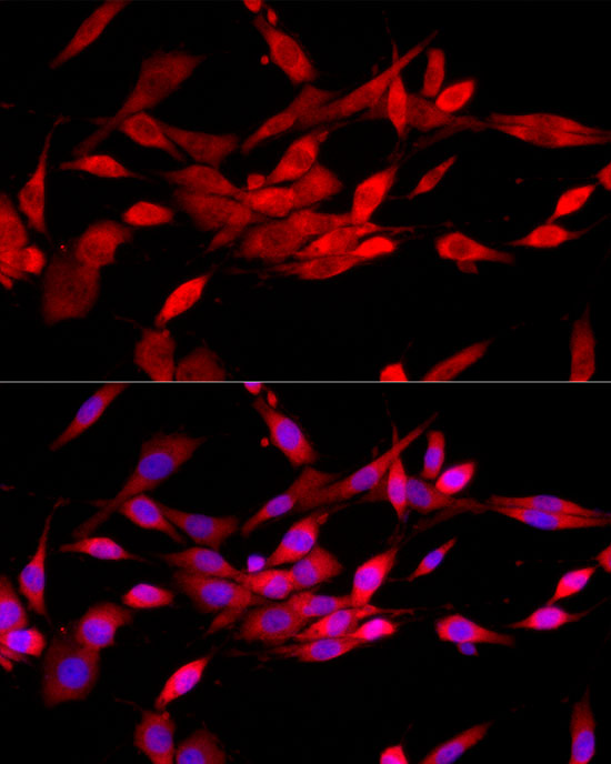
Immunofluorescence analysis of K-562 cells using CBL Rabbit pAb (A0732) at dilution of 1:300 (40x lens). Secondary antibody: Cy3-conjugated Goat anti-Rabbit IgG (H+L) (AS007) at 1:500 dilution. Blue: DAPI for nuclear staining. |
|
|
Immunofluorescence analysis of NIH/3T3 cells using CBL Rabbit pAb (A0732) at dilution of 1:300 (40x lens). Secondary antibody: Cy3-conjugated Goat anti-Rabbit IgG (H+L) (AS007) at 1:500 dilution. Blue: DAPI for nuclear staining. |
|
|
Immunofluorescence analysis of PC-12 cells using CBL Rabbit pAb (A0732) at dilution of 1:300 (40x lens). Secondary antibody: Cy3-conjugated Goat anti-Rabbit IgG (H+L) (AS007) at 1:500 dilution. Blue: DAPI for nuclear staining. |
|
|
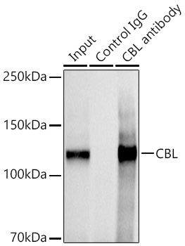
Immunoprecipitation analysis of 600 µg extracts of Mouse thymus cells using 3 µg CBL antibody (A0732). Western blot was performed from the immunoprecipitate using CBL antibody at a dilution of 1:1000. |
Product Guarantee and Expert Support
