
ERK1 / ERK2 Rabbit pAb, Unconjugated, Polyclonal
Catalog Number:
ABB-A16686
- Images (8)
| Article Name: | ERK1 / ERK2 Rabbit pAb, Unconjugated, Polyclonal |
| Biozol Catalog Number: | ABB-A16686 |
| Supplier Catalog Number: | A16686 |
| Alternative Catalog Number: | ABB-A16686-100UL,ABB-A16686-20UL,ABB-A16686-500UL,ABB-A16686-1000UL |
| Manufacturer: | ABclonal |
| Host: | Rabbit |
| Category: | Antikörper |
| Application: | ELISA, IF, IHC-P, WB |
| Species Reactivity: | Human |
| Immunogen: | Synthetic peptide. This information is considered to be commercially sensitive. |
| Conjugation: | Unconjugated |
| Alternative Names: | MAPK1/MAPK3, ERK1 / ERK2 |
| MAP kinases, also known as extracellular signal-regulated kinases (ERKs), act as an integration point for multiple biochemical signals, and are involved in a wide variety of cellular processes such as proliferation, differentiation, transcription regulation and development. The activation of this kinase requires its phosphorylation by upstream kinases. Upon activation, this kinase translocates to the nucleus of the stimulated cells, where it phosphorylates nuclear targets. |
| Application Dilute: | WB,1:500 - 1:5000|IF/ICC,1:50 - 1:200|IHC-P,1:50 - 1:200|ELISA,Recommended starting concentration is 1 µg/mL. Please optimize the concentration based on your specific assay requirements. |
| Application Notes: | Cross-Reactivity: Human,Mouse,Rat. ResearchArea: Protein phosphorylation. Shipping: Ice Bag |
|
|
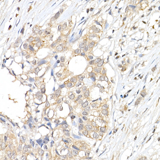
Immunohistochemistry analysis of paraffin-embedded Human breast cancer using ERK1 / ERK2 Rabbit pAb (A16686) at dilution of 1:50 (40x lens). High pressure antigen retrieval performed with 0.01M Citrate buffer (pH 6.0) prior to IHC staining. |
|
|
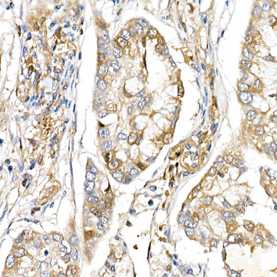
Immunohistochemistry analysis of paraffin-embedded Human lung cancer using ERK1 / ERK2 Rabbit pAb (A16686) at dilution of 1:50 (40x lens). High pressure antigen retrieval performed with 0.01M Citrate buffer (pH 6.0) prior to IHC staining. |
|
|
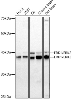
Western blot analysis of various lysates using ERK1 / ERK2 Rabbit pAb (A16686) at 1:1000 dilution. Secondary antibody: HRP-conjugated Goat anti-Rabbit IgG (H+L) (AS014) at 1:10000 dilution. Lysates/proteins: 25µg per lane. Blocking buffer: 3% nonfat dry milk in TBST. Detection: ECL Basic Kit (RM00020). Exposure time: 90s. |
|
|
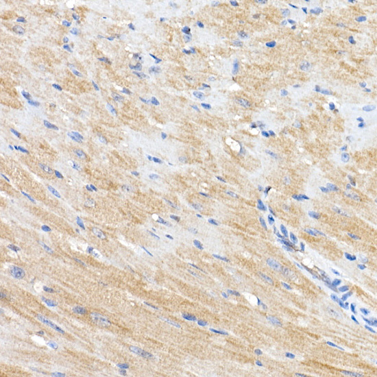
Immunohistochemistry analysis of paraffin-embedded Mouse heart using ERK1 / ERK2 Rabbit pAb (A16686) at dilution of 1:50 (40x lens). High pressure antigen retrieval performed with 0.01M Citrate buffer (pH 6.0) prior to IHC staining. |
|
|
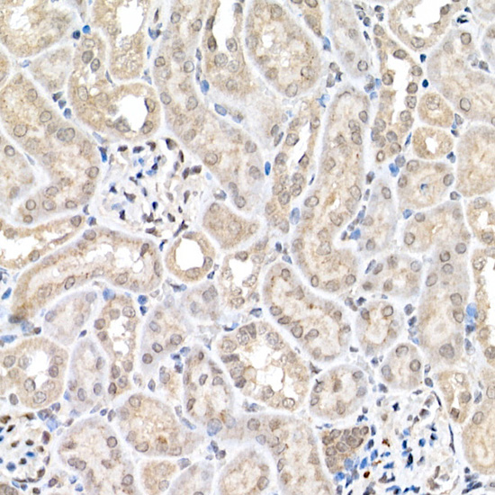
Immunohistochemistry analysis of paraffin-embedded Mouse kidney using ERK1 / ERK2 Rabbit pAb (A16686) at dilution of 1:50 (40x lens). High pressure antigen retrieval performed with 0.01M Citrate buffer (pH 6.0) prior to IHC staining. |
|
|
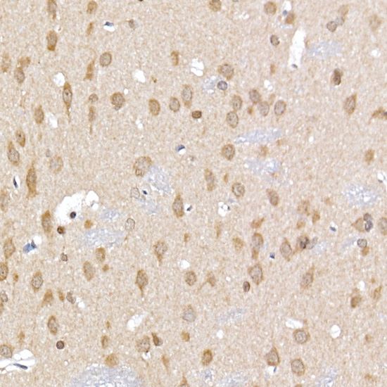
Immunohistochemistry analysis of paraffin-embedded Rat brain using ERK1 / ERK2 Rabbit pAb (A16686) at dilution of 1:50 (40x lens). High pressure antigen retrieval performed with 0.01M Citrate buffer (pH 6.0) prior to IHC staining. |
|
|
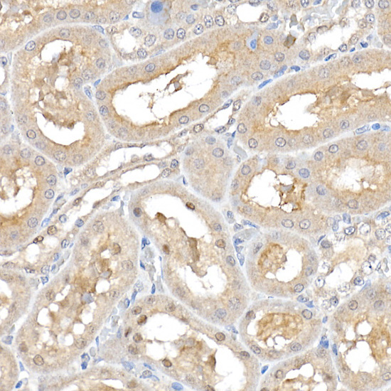
Immunohistochemistry analysis of paraffin-embedded Rat kidney using ERK1 / ERK2 Rabbit pAb (A16686) at dilution of 1:50 (40x lens). High pressure antigen retrieval performed with 0.01M Citrate buffer (pH 6.0) prior to IHC staining. |
|
|
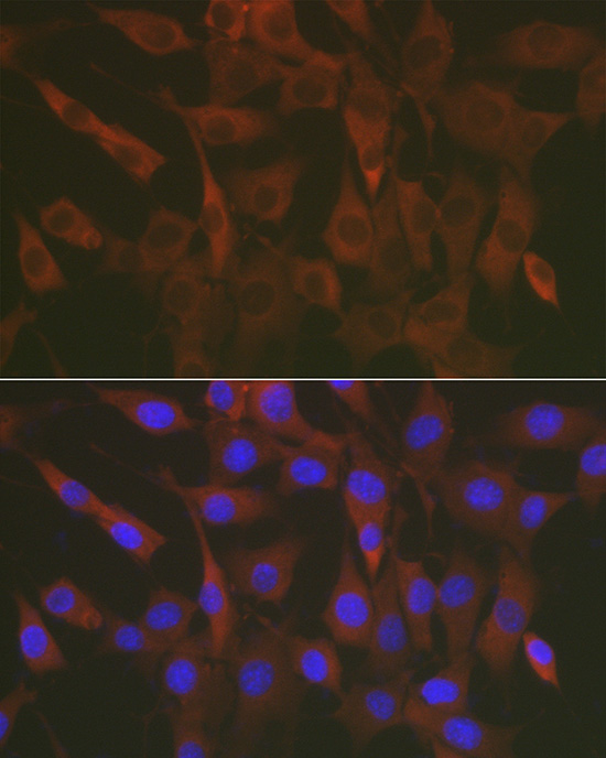
Immunofluorescence analysis of NIH/3T3 cells using ERK1 / ERK2 Rabbit pAb (A16686) at dilution of 1:50 (40x lens). Secondary antibody: Cy3-conjugated Goat anti-Rabbit IgG (H+L) (AS007) at 1:500 dilution. Blue: DAPI for nuclear staining. |
Product Guarantee and Expert Support
