
CD105/Endoglin Rabbit mAb, Unconjugated, Monoclonal
Catalog Number:
ABB-A19008
- Images (8)

| Article Name: | CD105/Endoglin Rabbit mAb, Unconjugated, Monoclonal |
| Biozol Catalog Number: | ABB-A19008 |
| Supplier Catalog Number: | A19008 |
| Alternative Catalog Number: | ABB-A19008-100UL,ABB-A19008-20UL,ABB-A19008-1000UL,ABB-A19008-500UL |
| Manufacturer: | ABclonal |
| Host: | Rabbit |
| Category: | Antikörper |
| Application: | ELISA, IHC-P, WB |
| Species Reactivity: | Human |
| Immunogen: | Recombinant protein (or fragment).This information is considered to be commercially sensitive. |
| Conjugation: | Unconjugated |
| Alternative Names: | END, HHT1, ORW1, CD105/Endoglin |
| This gene encodes a homodimeric transmembrane protein which is a major glycoprotein of the vascular endothelium. This protein is a component of the transforming growth factor beta receptor complex and it binds to the beta1 and beta3 peptides with high affinity. Mutations in this gene cause hereditary hemorrhagic telangiectasia, also known as Osler-Rendu-Weber syndrome 1, an autosomal dominant multisystemic vascular dysplasia. This gene may also be involved in preeclampsia and several types of cancer. Alternatively spliced transcript variants encoding different isoforms have been found for this gene. |
| Clonality: | Monoclonal |
| Clone Designation: | [ARC0446] |
| Molecular Weight: | 71kDa |
| NCBI: | 2022 |
| UniProt: | P17813 |
| Purity: | Affinity purification |
| Sequence: | MDRGTLPLAVALLLASCSLSPTSLAETVHCDLQPVGPERGEVTYTTSQVSKGCVAQAPNAILEVHVLFLEFPTGPSQLELTLQASKQNGTWPREVLLVLSVNSSVFLHLQALGIPLHLAYNSSLVTFQEPPGVNTTELPSFPKTQILEWAAERGPITSAAELNDPQSILLRLGQAQGSLSFCMLEASQDMGRTLEWRPRT |
| Target: | ENG |
| Antibody Type: | Primary Antibody |
| Application Dilute: | WB,1:1000 - 1:2000|IHC-P,1:5000 - 1:20000|ELISA,Recommended starting concentration is 1 µg/mL. Please optimize the concentration based on your specific assay requirements. |
| Application Notes: | Cross-Reactivity: Human. ResearchArea: Cancer,Tumor biomarkers,Immunology Inflammation,CDs,Stem Cells,Mesenchymal Stem Cells,Cardiovascular. Shipping: Ice Bag |
|
|
Western blot analysis of various lysates using CD105/Endoglin Rabbit mAb (A19008) at 1:1000 dilution incubated overnight at 4°C. Secondary antibody: HRP-conjugated Goat anti-Rabbit IgG (H+L) (AS014) at 1:10000 dilution. Lysates/proteins: 25 µg per lane. Blocking buffer: 3% nonfat dry milk in TBST. Detection: ECL Basic Kit (RM00020). Exposure time: 20s. |
|
|
Western blot analysis of lysates from HeLa cells using CD105/Endoglin Rabbit mAb (A19008) at 1:1000 dilutionincubated overnight at 4°C. HeLa cells were treated with PNGase (1 µL) at 37°C for 3 hours. Secondary antibody: HRP-conjugated Goat anti-Rabbit IgG (H+L) (AS014) at 1:10000 dilution. Lysates/proteins: 30 µg per lane. Blocking buffer: 3 % nonfat dry milk in TBST. Detection: ECL Basic Kit (RM00020). Exposure time: 60s. |
|
|
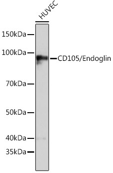
Western blot analysis of lysates from HUVEC cells, using CD105/Endoglin Rabbit mAb (A19008) at 1:1000 dilution. Secondary antibody: HRP-conjugated Goat anti-Rabbit IgG (H+L) (AS014) at 1:10000 dilution. Lysates/proteins: 25µg per lane. Blocking buffer: 3% nonfat dry milk in TBST. Detection: ECL Basic Kit (RM00020). Exposure time: 60s. |
|
|
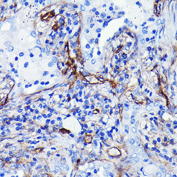
Immunohistochemistry analysis of paraffin-embedded Human liver tissue using CD105/Endoglin Rabbit mAb (A19008) at a dilution of 1:10000 (40x lens). High pressure antigen retrieval performed with 0.01M Tris-EDTA Buffer (pH 9.0) prior to IHC staining. |
|
|
Immunohistochemistry analysis of paraffin-embedded Human breast cancer tissue using CD105/Endoglin Rabbit mAb (A19008) at a dilution of 1:10000 (40x lens). High pressure antigen retrieval performed with 0.01M Tris-EDTA Buffer (pH 9.0) prior to IHC staining. |
|
|
Immunohistochemistry analysis of paraffin-embedded Human placenta tissue using CD105/Endoglin Rabbit mAb (A19008) at a dilution of 1:10000 (40x lens). High pressure antigen retrieval performed with 0.01M Tris-EDTA Buffer (pH 9.0) prior to IHC staining. |
|
|
Immunohistochemistry analysis of paraffin-embedded Human prostate tissue using CD105/Endoglin Rabbit mAb (A19008) at a dilution of 1:10000 (40x lens). High pressure antigen retrieval performed with 0.01M Tris-EDTA Buffer (pH 9.0) prior to IHC staining. |
|
|
Immunohistochemistry analysis of paraffin-embedded Human tonsil tissue using CD105/Endoglin Rabbit mAb (A19008) at a dilution of 1:10000 (40x lens). High pressure antigen retrieval performed with 0.01M Tris-EDTA Buffer (pH 9.0) prior to IHC staining. |
Product Guarantee and Expert Support
