
[KD Validated] ATG7 Rabbit mAb, Unconjugated, Monoclonal
Catalog Number:
ABB-A19604
- Images (8)

| Article Name: | [KD Validated] ATG7 Rabbit mAb, Unconjugated, Monoclonal |
| Biozol Catalog Number: | ABB-A19604 |
| Supplier Catalog Number: | A19604 |
| Alternative Catalog Number: | ABB-A19604-20UL,ABB-A19604-100UL,ABB-A19604-1000UL,ABB-A19604-500UL |
| Manufacturer: | ABclonal |
| Host: | Rabbit |
| Category: | Antikörper |
| Application: | ELISA, IHC-P, IP, WB |
| Species Reactivity: | Human |
| Immunogen: | Synthetic peptide. This information is considered to be commercially sensitive. |
| Conjugation: | Unconjugated |
| Alternative Names: | GSA7, [KD Validated] ATG7 |
| This gene encodes an E1-like activating enzyme that is essential for autophagy and cytoplasmic to vacuole transport. The encoded protein is also thought to modulate p53-dependent cell cycle pathways during prolonged metabolic stress. It has been associated with multiple functions, including axon membrane trafficking, axonal homeostasis, mitophagy, adipose differentiation, and hematopoietic stem cell maintenance. Alternative splicing results in multiple transcript variants. |
| Application Dilute: | WB,1:1000 - 1:6000|IHC-P,1:200 - 1:800|IP,0.5µg-4µg antibody for 200µg-400µg extracts of whole cells|ELISA,Recommended starting concentration is 1 µg/mL. Please optimize the concentration based on your specific assay requirements. |
| Application Notes: | Cross-Reactivity: Human,Mouse,Rat. ResearchArea: Cancer,Signal Transduction,Cell Biology Developmental Biology,Apoptosis,Autophagy,Ubiquitin,Endocrine Metabolism,Mitochondrial metabolism,Immunology Inflammation,Cardiovascular,Heart. Shipping: Ice Bag |
|
|
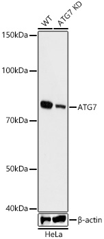
Western blot analysis of lysates from wild type(WT) and ATG7 knockdown (KD) HeLa cells using [KD Validated] ATG7 Rabbit mAb (A19604) at 1:1000 dilution incubated overnight at 4°C. Secondary antibody: HRP-conjugated Goat anti-Rabbit IgG (H+L) (AS014) at 1:10000 dilution. Lysates/proteins: 25µg per lane. Blocking buffer: 3% nonfat dry milk in TBST. Detection: ECL Basic Kit (RM00020). Exposure time: 60s. |
|
|
Immunohistochemistry analysis of paraffin-embedded Mouse brain tissue using [KD Validated] ATG7 Rabbit mAb (A19604) at a dilution of 1:500 (40x lens). High pressure antigen retrieval performed with 0.01M Tris-EDTA Buffer (pH 9.0) prior to IHC staining. |
|
|
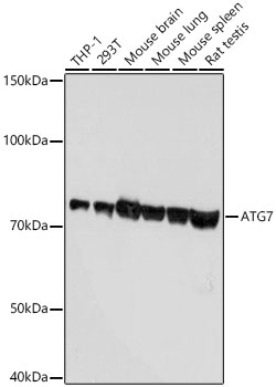
Western blot analysis of various lysates using [KD Validated] ATG7 Rabbit mAb (A19604) at 1:1000 dilution incubated overnight at 4°C. Secondary antibody: HRP-conjugated Goat anti-Rabbit IgG (H+L) (AS014) at 1:10000 dilution. Lysates/proteins: 25µg per lane. Blocking buffer: 3% nonfat dry milk in TBST. Detection: ECL Basic Kit (RM00020). Exposure time: 10s. |
|
|
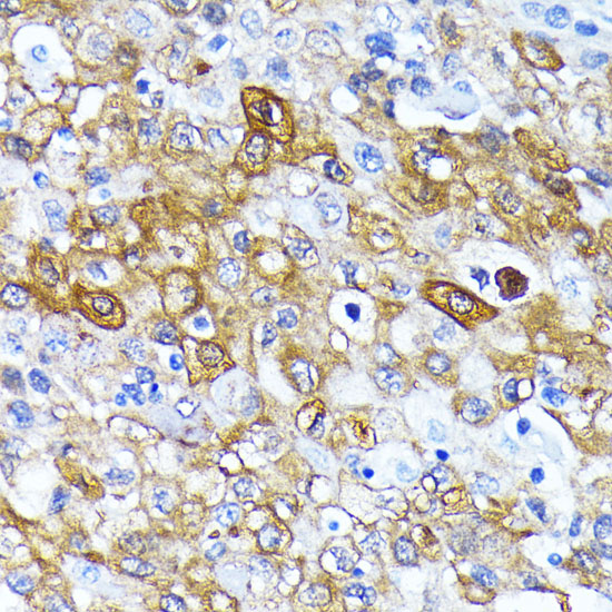
Immunohistochemistry analysis of paraffin-embedded Human lung cancer tissue using [KD Validated] ATG7 Rabbit mAb (A19604) at a dilution of 1:500 (40x lens). High pressure antigen retrieval performed with 0.01M Tris-EDTA Buffer (pH 9.0) prior to IHC staining. |
|
|
Immunohistochemistry analysis of paraffin-embedded Mouse testis tissue using [KD Validated] ATG7 Rabbit mAb (A19604) at a dilution of 1:500 (40x lens). High pressure antigen retrieval performed with 0.01M Tris-EDTA Buffer (pH 9.0) prior to IHC staining. |
|
|
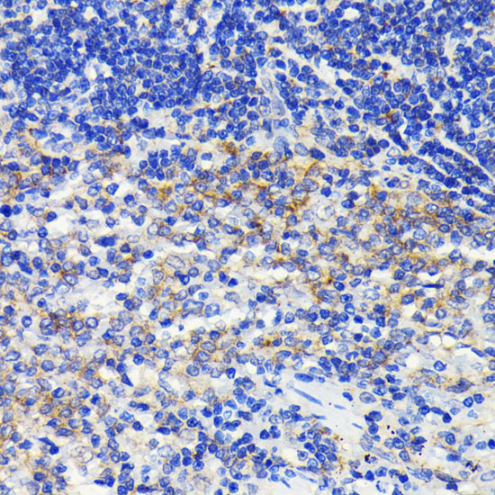
Immunohistochemistry analysis of paraffin-embedded Rat colon tissue using [KD Validated] ATG7 Rabbit mAb (A19604) at a dilution of 1:500 (40x lens). High pressure antigen retrieval performed with 0.01M Tris-EDTA Buffer (pH 9.0) prior to IHC staining. |
|
|
Immunohistochemistry analysis of paraffin-embedded Rat testis tissue using [KD Validated] ATG7 Rabbit mAb (A19604) at a dilution of 1:500 (40x lens). High pressure antigen retrieval performed with 0.01M Tris-EDTA Buffer (pH 9.0) prior to IHC staining. |
|
|
Immunoprecipitation of ATG7 from 300 µg extracts of HeLa cells was performed using 2 µg of [KD Validated] ATG7 Rabbit mAb (A19604). Rabbit IgG isotype control (AC005) was used to precipitate the Control IgG sample. IP samples were eluted with 1X reducing Laemmli Buffer. The Input lane represents 10% of the total input. Western blot analysis of immunoprecipitates was conducted using [KD Validated] ATG7 Rabbit mAb (A19604) at a dilution of 1:1000. |
Product Guarantee and Expert Support
