
PSMA/FOLH1 Rabbit mAb, Unconjugated, Monoclonal
Catalog Number:
ABB-A22212
- Images (8)

| Article Name: | PSMA/FOLH1 Rabbit mAb, Unconjugated, Monoclonal |
| Biozol Catalog Number: | ABB-A22212 |
| Supplier Catalog Number: | A22212 |
| Alternative Catalog Number: | ABB-A22212-20UL,ABB-A22212-100UL |
| Manufacturer: | ABclonal |
| Host: | Rabbit |
| Category: | Antikörper |
| Application: | ELISA, IHC-P, WB |
| Species Reactivity: | Human |
| Immunogen: | Synthetic peptide. This information is considered to be commercially sensitive. |
| Conjugation: | Unconjugated |
| Alternative Names: | PSM, FGCP, FOLH, GCP2, PSMA, mGCP, GCPII, NAALAD1, PSMA/FOLH1 |
| This gene encodes a type II transmembrane glycoprotein belonging to the M28 peptidase family. The protein acts as a glutamate carboxypeptidase on different alternative substrates, including the nutrient folate and the neuropeptide N-acetyl-l-aspartyl-l-glutamate and is expressed in a number of tissues such as prostate, central and peripheral nervous system and kidney. A mutation in this gene may be associated with impaired intestinal absorption of dietary folates, resulting in low blood folate levels and consequent hyperhomocysteinemia. Expression of this protein in the brain may be involved in a number of pathological conditions associated with glutamate excitotoxicity. In the prostate the protein is up-regulated in cancerous cells and is used as an effective diagnostic and prognostic indicator of prostate cancer. This gene likely arose from a duplication event of a nearby chromosomal region. Alternative splicing gives rise to multiple transcript variants encoding several different isoforms. |
| Application Dilute: | WB,1:20000 - 1:120000|IHC-P,1:5000 - 1:20000|ELISA,Recommended starting concentration is 1 µg/mL. Please optimize the concentration based on your specific assay requirements. |
| Application Notes: | Cross-Reactivity: Human,Mouse,Rat. ResearchArea: Cancer,Tumor biomarkers,Signal Transduction,Endocrine Metabolism,Amino acid metabolism. Shipping: Ice Bag |
|
|
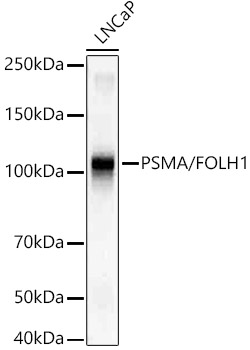
Western blot analysis of lysates from LNCaP cells, using PSMA/FOLH1 Rabbit mAb (A22212) at1:20000 dilution. Secondary antibody: HRP-conjugated Goat anti-Rabbit IgG (H+L) (AS014) at 1:10000 dilution. Lysates/proteins: 25µg per lane. Blocking buffer: 3% nonfat dry milk in TBST. Detection: ECL Basic Kit (RM00020). Exposure time: 1s. |
|
|
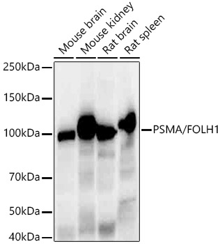
Western blot analysis of various lysates, using PSMA/FOLH1 Rabbit mAb (A22212) at1:20000 dilution. Secondary antibody: HRP-conjugated Goat anti-Rabbit IgG (H+L) (AS014) at 1:10000 dilution. Lysates/proteins: 25µg per lane. Blocking buffer: 3% nonfat dry milk in TBST. Detection: ECL Enhanced Kit (RM00021). Exposure time: 30s. |
|
|
Immunohistochemistry analysis of paraffin-embedded Human cervix cancer tissue using PSMA/FOLH1 Rabbit mAb (A22212) at a dilution of 1:15000 (40x lens). High pressure antigen retrieval performed with 0.01M Tris-EDTA Buffer (pH 9.0) prior to IHC staining. |
|
|
Immunohistochemistry analysis of paraffin-embedded Human colon carcinoma tissue using PSMA/FOLH1 Rabbit mAb (A22212) at a dilution of 1:15000 (40x lens). High pressure antigen retrieval performed with 0.01M Tris-EDTA Buffer (pH 9.0) prior to IHC staining. |
|
|
Immunohistochemistry analysis of paraffin-embedded Human kidney tissue using PSMA/FOLH1 Rabbit mAb (A22212) at a dilution of 1:15000 (40x lens). High pressure antigen retrieval performed with 0.01M Tris-EDTA Buffer (pH 9.0) prior to IHC staining. |
|
|
Immunohistochemistry analysis of paraffin-embedded Human prostate cancer tissue using PSMA/FOLH1 Rabbit mAb (A22212) at a dilution of 1:15000 (40x lens). High pressure antigen retrieval performed with 0.01M Tris-EDTA Buffer (pH 9.0) prior to IHC staining. |
|
|
Immunohistochemistry analysis of paraffin-embedded Mouse kidney tissue using PSMA/FOLH1 Rabbit mAb (A22212) at a dilution of 1:15000 (40x lens). High pressure antigen retrieval performed with 0.01M Tris-EDTA Buffer (pH 9.0) prior to IHC staining. |
|
|
Confocal imaging of LNCaP cells using PSMA/FOLH1 Rabbit mAb (A22212, dilution 1:200) followed by a further incubation with Cy3-conjugated Goat anti-Rabbit IgG (H+L) (AS007, dilution 1:500) (Red). DAPI was used for nuclear staining (Blue). Objective: 100x. |
Product Guarantee and Expert Support
