
TriMethyl-Histone H3-K4 Rabbit mAb, Unconjugated
Catalog Number:
ABB-A22224
- Images (8)
| Article Name: | TriMethyl-Histone H3-K4 Rabbit mAb, Unconjugated |
| Biozol Catalog Number: | ABB-A22224 |
| Supplier Catalog Number: | A22224 |
| Alternative Catalog Number: | ABB-A22224-20UL,ABB-A22224-100UL,ABB-A22224-1000UL,ABB-A22224-500UL |
| Manufacturer: | ABclonal |
| Host: | Rabbit |
| Category: | Antikörper |
| Application: | ChIP, ELISA, IF, IHC-P, WB |
| Species Reactivity: | Human |
| Immunogen: | Synthetic peptide. This information is considered to be commercially sensitive. |
| Conjugation: | Unconjugated |
| Alternative Names: | H3.4, H3/g, H3FT, H3t, HIST3H3, Histone H3, HIST1H3A, TriMethyl-Histone H3-K4 |
| Histones are basic nuclear proteins that are responsible for the nucleosome structure of the chromosomal fiber in eukaryotes. Nucleosomes consist of approximately 146 bp of DNA wrapped around a histone octamer composed of pairs of each of the four core histones (H2A, H2B, H3, and H4). The chromatin fiber is further compacted through the interaction of a linker histone, H1, with the DNA between the nucleosomes to form higher order chromatin structures. This gene is intronless and encodes a replication-dependent histone that is a member of the histone H3 family. Transcripts from this gene lack polyA tails, instead, they contain a palindromic termination element. This gene is located separately from the other H3 genes that are in the histone gene cluster on chromosome 6p22-p21.3. |
| Application Dilute: | WB,1:10000 - 1:30000|IHC-P,1:50 - 1:200|IF/ICC,1:50 - 1:200|ELISA,Recommended starting concentration is 1 µg/mL. Please optimize the concentration based on your specific assay requirements.|ChIP,5µg antibody for 5µg-10µg of Chromatin |
| Application Notes: | Cross-Reactivity: Human,Mouse,Rat,Other (Wide Range Predicted). ResearchArea: Signal Transduction,MAPK-Erk Signaling Pathway,MAPK-P38 Signaling Pathway,Epigenetics & Nuclear Signaling,Epigenetic Modifications,Methylation. Shipping: Ice Bag |
|
|
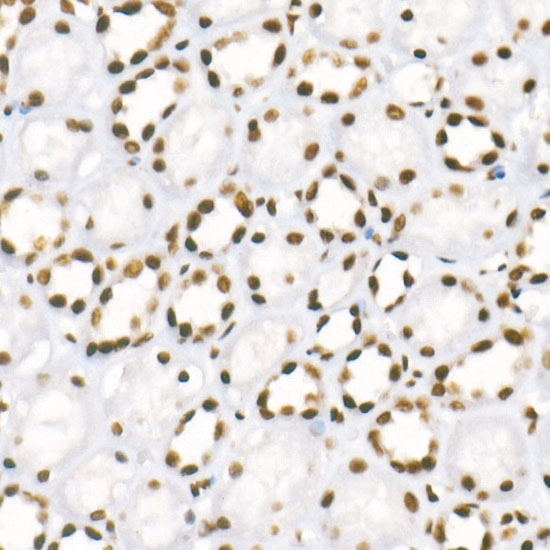
Immunohistochemistry analysis of paraffin-embedded Human kidney using TriMethyl-Histone H3-K4 Rabbit mAb (A22224) at dilution of 1:100 (40x lens). High pressure antigen retrieval performed with 0.01M Citrate buffer (pH 6.0) prior to IHC staining. |
|
|
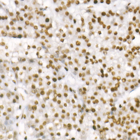
Immunohistochemistry analysis of paraffin-embedded Human liver cancer using TriMethyl-Histone H3-K4 Rabbit mAb (A22224) at dilution of 1:100 (40x lens). High pressure antigen retrieval performed with 0.01M Citrate buffer (pH 6.0) prior to IHC staining. |
|
|
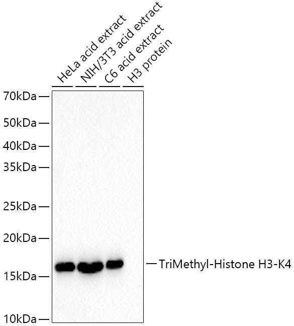
Western blot analysis of various lysates, using TriMethyl-Histone H3-K4 Rabbit mAb (A22224) at 1:30000 dilution. Secondary antibody: HRP-conjugated Goat anti-Rabbit IgG (H+L) (AS014) at 1:10000 dilution. Lysates/proteins: 25µg per lane. Blocking buffer: 3% nonfat dry milk in TBST. Detection: ECL Basic Kit (RM00020). Exposure time: 30s. |
|
|
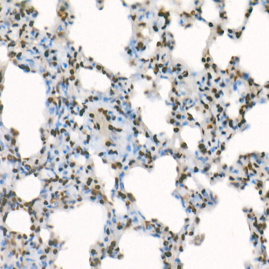
Immunohistochemistry analysis of paraffin-embedded Rat kidney using TriMethyl-Histone H3-K4 Rabbit mAb (A22224) at dilution of 1:100 (40x lens). High pressure antigen retrieval performed with 0.01M Citrate buffer (pH 6.0) prior to IHC staining. |
|
|
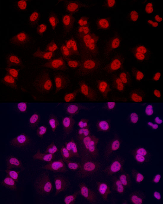
Immunofluorescence analysis of C6 cells using TriMethyl-Histone H3-K4 Rabbit mAb (A22224) at dilution of 1:100(40x lens). Secondary antibody: Cy3-conjugated Goat anti-Rabbit IgG (H+L) (AS007) at 1:500 dilution. Blue: DAPI for nuclear staining. |
|
|
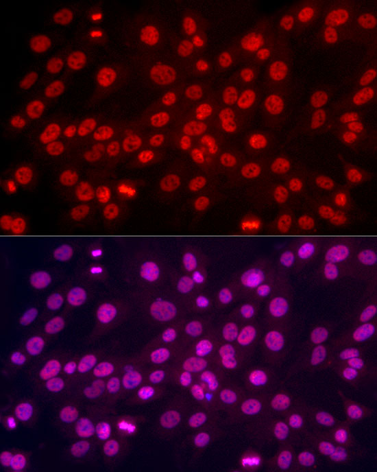
Immunofluorescence analysis of HeLa cells using TriMethyl-Histone H3-K4 Rabbit mAb (A22224) at dilution of 1:100(40x lens). Secondary antibody: Cy3-conjugated Goat anti-Rabbit IgG (H+L) (AS007) at 1:500 dilution. Blue: DAPI for nuclear staining. |
|
|
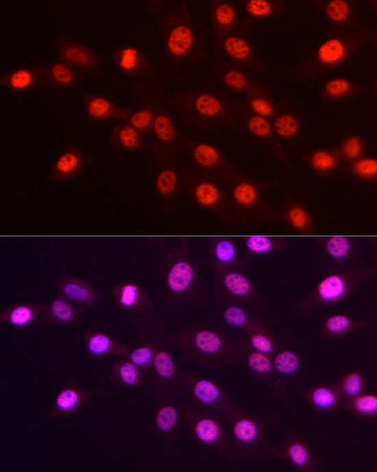
Immunofluorescence analysis of NIH/3T3 cells using TriMethyl-Histone H3-K4 Rabbit mAb (A22224) at dilution of 1:100(40x lens). Secondary antibody: Cy3-conjugated Goat anti-Rabbit IgG (H+L) (AS007) at 1:500 dilution. Blue: DAPI for nuclear staining. |
|
|
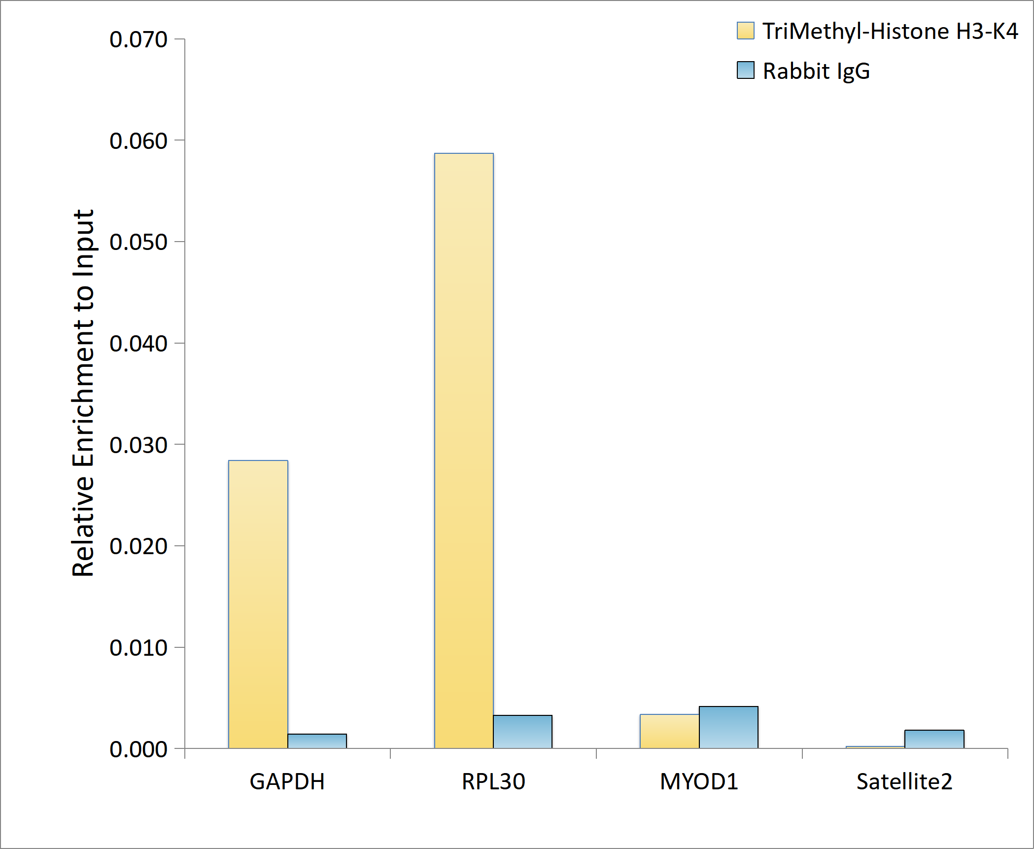
Chromatin immunoprecipitation analysis of extracts of HeLa cells, using TriMethyl-Histone H3-K4 antibody (A22224) and rabbit IgG.The amount of immunoprecipitated DNA was checked by quantitative PCR. Histogram was constructed by the ratios of the immunoprecipitated DNA to the input. |
Product Guarantee and Expert Support
