
RbAp46/RBBP7 Rabbit mAb, Unconjugated, Monoclonal
Catalog Number:
ABB-A22274
- Images (8)

| Article Name: | RbAp46/RBBP7 Rabbit mAb, Unconjugated, Monoclonal |
| Biozol Catalog Number: | ABB-A22274 |
| Supplier Catalog Number: | A22274 |
| Alternative Catalog Number: | ABB-A22274-100UL,ABB-A22274-20UL |
| Manufacturer: | ABclonal |
| Host: | Rabbit |
| Category: | Antikörper |
| Application: | ELISA, IF, IHC-P, WB |
| Species Reactivity: | Human |
| Immunogen: | Recombinant protein (or fragment).This information is considered to be commercially sensitive. |
| Conjugation: | Unconjugated |
| Alternative Names: | RbAp46, RbAp46/RBBP7 |
| This protein is a ubiquitously expressed nuclear protein and belongs to a highly conserved subfamily of WD-repeat proteins. It is found among several proteins that binds directly to retinoblastoma protein, which regulates cell proliferation. The encoded protein is found in many histone deacetylase complexes, including mSin3 co-repressor complex. It is also present in protein complexes involved in chromatin assembly. This protein can interact with BRCA1 tumor-suppressor gene and may have a role in the regulation of cell proliferation and differentiation. Two transcript variants encoding different isoforms have been found for this gene. |
| Application Dilute: | WB,1:2000 - 1:20000|IHC-P,1:100 - 1:500|IF/ICC,1:50 - 1:200|ELISA,Recommended starting concentration is 1 µg/mL. Please optimize the concentration based on your specific assay requirements. |
| Application Notes: | Cross-Reactivity: Human,Mouse,Rat. ResearchArea: Epigenetics Nuclear Signaling,Chromatin Remodeling,Cell Biology Developmental Biology,Cell Cycle. Shipping: Ice Bag |
|
|
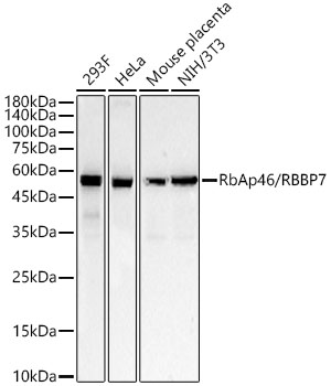
Western blot analysis of various lysates, using RbAp46/RBBP7 Rabbit mAb (A22274) at 1:20000 dilution. Secondary antibody: HRP-conjugated Goat anti-Rabbit IgG (H+L) (AS014) at 1:10000 dilution. Lysates/proteins: 25µg per lane. Blocking buffer: 3% nonfat dry milk in TBST. Detection: ECL Basic Kit (RM00020). Exposure time: 20s. |
|
|
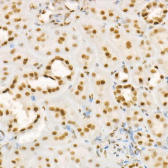
Immunohistochemistry analysis of paraffin-embeddedRat liver tissue usingRbAp46/RBBP7 Rabbit mAb(A22274) at a dilution of 1:400 (40x lens).High pressure antigen retrieval was performed with 0.01 M citrate buffer (pH 6.0) prior to IHC staining. |
|
|
Immunohistochemistry analysis of paraffin-embeddedMouse colon tissue usingRbAp46/RBBP7 Rabbit mAb(A22274) at a dilution of 1:400 (40x lens).High pressure antigen retrieval was performed with 0.01 M citrate buffer (pH 6.0) prior to IHC staining. |
|
|
Immunohistochemistry analysis of paraffin-embeddedMouse testis tissue usingRbAp46/RBBP7 Rabbit mAb(A22274) at a dilution of 1:400 (40x lens).High pressure antigen retrieval was performed with 0.01 M citrate buffer (pH 6.0) prior to IHC staining. |
|
|
Immunohistochemistry analysis of paraffin-embeddedRat colon tissue usingRbAp46/RBBP7 Rabbit mAb(A22274) at a dilution of 1:400 (40x lens).High pressure antigen retrieval was performed with 0.01 M citrate buffer (pH 6.0) prior to IHC staining. |
|
|
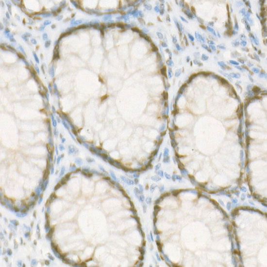
Immunohistochemistry analysis of paraffin-embeddedHuman small intestine tissue usingRbAp46/RBBP7 Rabbit mAb(A22274) at a dilution of 1:400 (40x lens).High pressure antigen retrieval was performed with 0.01 M citrate buffer (pH 6.0) prior to IHC staining. |
|
|
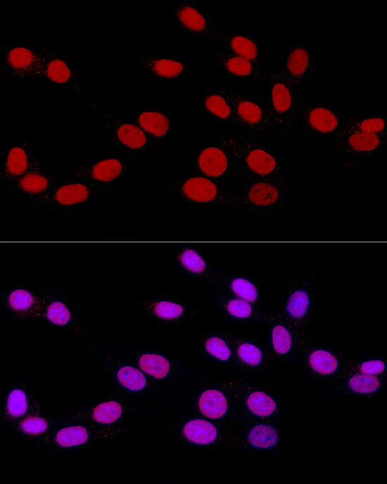
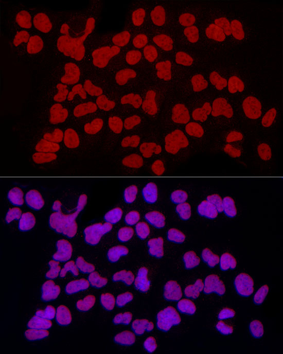
Immunofluorescence analysis of NIH/3T3 cells using RbAp46/RBBP7 Rabbit mAb (A22274) at dilution of 1:100(40x lens). Secondary antibody: Cy3-conjugated Goat anti-Rabbit IgG (H+L) (AS007) at 1:500 dilution. Blue: DAPI for nuclear staining. |
|
|
Immunofluorescence analysis of U-2 OS cells using RbAp46/RBBP7 Rabbit mAb (A22274) at dilution of 1:100(40x lens). Secondary antibody: Cy3-conjugated Goat anti-Rabbit IgG (H+L) (AS007) at 1:500 dilution. Blue: DAPI for nuclear staining. |
Product Guarantee and Expert Support
