
KAT2B/PCAF Rabbit mAb, Unconjugated, Monoclonal
Catalog Number:
ABB-A22719
- Images (8)


| Article Name: | KAT2B/PCAF Rabbit mAb, Unconjugated, Monoclonal |
| Biozol Catalog Number: | ABB-A22719 |
| Supplier Catalog Number: | A22719 |
| Alternative Catalog Number: | ABB-A22719-100UL,ABB-A22719-20UL |
| Manufacturer: | ABclonal |
| Host: | Rabbit |
| Category: | Antikörper |
| Application: | ELISA, IHC-P, IP, WB |
| Species Reactivity: | Human |
| Immunogen: | Recombinant protein (or fragment).This information is considered to be commercially sensitive. |
| Conjugation: | Unconjugated |
| Alternative Names: | CAF, PCAF, P/CAF, KAT2B/PCAF |
| CBP and p300 are large nuclear proteins that bind to many sequence-specific factors involved in cell growth and/or differentiation, including c-jun and the adenoviral oncoprotein E1A. The protein encoded by this gene associates with p300/CBP. It has in vitro and in vivo binding activity with CBP and p300, and competes with E1A for binding sites in p300/CBP. It has histone acetyl transferase activity with core histones and nucleosome core particles, indicating that this protein plays a direct role in transcriptional regulation. |
| Application Dilute: | WB,1:500 - 1:2000|IP,0.5µg-4µg antibody for 400µg-600µg extracts of whole cells|IHC-P,1:200 - 1:2000|ELISA,Recommended starting concentration is 1 µg/mL. Please optimize the concentration based on your specific assay requirements. |
| Application Notes: | Cross-Reactivity: Human,Mouse,Rat. ResearchArea: Epigenetics Nuclear Signaling,Epigenetic writers and erasers of core Histones,Nuclear Receptor Signaling,Signal Transduction,Cell Biology Developmental Biology,Cell Cycle,Cell cycle inhibitors,Cell Cycle Control-G2 M DNA Damage Checkpoint,Immunology Inflammation,NF-kB Signaling Pathway. Shipping: Ice Bag |
|
|
Western blot analysis of various lysates using KAT2B/PCAF Rabbit mAb (A22719) at 1:1000 dilution incubated at room temperature for 1.5 hours. Secondary antibody: HRP-conjugated Goat anti-Rabbit IgG (H+L) (AS014) at 1:10000 dilution. Lysates/proteins: 25 µg per lane. Blocking buffer: 3% nonfat dry milk in TBST. Detection: ECL Basic Kit (RM00020). Exposure time: 90s. |
|
|
Immunohistochemistry analysis of paraffin-embeddedRat colon tissue usingKAT2B/PCAF Rabbit mAb(A22719) at a dilution of 1:200 (40x lens).High pressure antigen retrieval was performed with 0.01 M citrate buffer (pH 6.0) prior to IHC staining. |
|
|
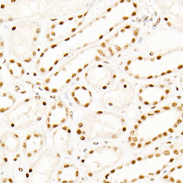
Immunohistochemistry analysis of paraffin-embeddedRat kidney tissue usingKAT2B/PCAF Rabbit mAb(A22719) at a dilution of 1:200 (40x lens).High pressure antigen retrieval was performed with 0.01 M citrate buffer (pH 6.0) prior to IHC staining. |
|
|
Immunohistochemistry analysis of paraffin-embeddedMouse colon tissue usingKAT2B/PCAF Rabbit mAb(A22719) at a dilution of 1:200 (40x lens).High pressure antigen retrieval was performed with 0.01 M citrate buffer (pH 6.0) prior to IHC staining. |
|
|
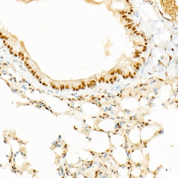
Immunohistochemistry analysis of paraffin-embeddedMouse kidney tissue usingKAT2B/PCAF Rabbit mAb(A22719) at a dilution of 1:200 (40x lens).High pressure antigen retrieval was performed with 0.01 M citrate buffer (pH 6.0) prior to IHC staining. |
|
|
Immunohistochemistry analysis of paraffin-embeddedHuman pancreas tissue usingKAT2B/PCAF Rabbit mAb(A22719) at a dilution of 1:200 (40x lens).High pressure antigen retrieval was performed with 0.01 M citrate buffer (pH 6.0) prior to IHC staining. |
|
|
Immunohistochemistry analysis of paraffin-embeddedHuman thyroid tissue usingKAT2B/PCAF Rabbit mAb(A22719) at a dilution of 1:200 (40x lens).High pressure antigen retrieval was performed with 0.01 M citrate buffer (pH 6.0) prior to IHC staining. |
|
|
Immunoprecipitation of KAT2B/PCAF from 600 µg extracts of Hep G2 cells was performed using 2 µg of KAT2B/PCAF Rabbit mAb (A22719). Rabbit Control IgG (AC005) was used to precipitate the Control IgG sample. IP samples were eluted with 1X Laemmli Buffer. The Input lane represents 10% of the total input. Western blot analysis of immunoprecipitates was conducted using KAT2B/PCAF Rabbit mAb (A22719) at a dilution of 1:1000. |
Product Guarantee and Expert Support
