
c-Fos Rabbit mAb, Unconjugated, Monoclonal
Catalog Number:
ABB-A24620
- Images (8)

| Article Name: | c-Fos Rabbit mAb, Unconjugated, Monoclonal |
| Biozol Catalog Number: | ABB-A24620 |
| Supplier Catalog Number: | A24620 |
| Alternative Catalog Number: | ABB-A24620-20UL,ABB-A24620-100UL,ABB-A24620-1000UL,ABB-A24620-500UL |
| Manufacturer: | ABclonal |
| Host: | Rabbit |
| Category: | Antikörper |
| Application: | ELISA, IF, IHC-P, WB |
| Species Reactivity: | Human |
| Immunogen: | Recombinant protein (or fragment).This information is considered to be commercially sensitive. |
| Conjugation: | Unconjugated |
| Alternative Names: | p55, AP-1, C-FOS, c-Fos |
| The Fos gene family consists of 4 members: FOS, FOSB, FOSL1, and FOSL2. These genes encode leucine zipper proteins that can dimerize with proteins of the JUN family, thereby forming the transcription factor complex AP-1. As such, the FOS proteins have been implicated as regulators of cell proliferation, differentiation, and transformation. In some cases, expression of the FOS gene has also been associated with apoptotic cell death. |
| Application Dilute: | WB,1:1000 - 1:4000|IF-P,1:200 - 1:400|IHC-P,1:200 - 1:800|ELISA,Recommended starting concentration is 1 µg/mL. Please optimize the concentration based on your specific assay requirements. |
| Application Notes: | Cross-Reactivity: Human,Mouse,Rat. ResearchArea: Epigenetics Nuclear Signaling,Transcription Factors,Protein phosphorylation,Cancer,Tumor biomarkers,Signal Transduction,ErbB-HER Signaling Pathway,MAPK-Erk Signaling Pathway,Immunology Inflammation,T Cell Receptor Signaling Pathway,Neuroscience, Cell Type Marker,Neuron marker. Shipping: Ice Bag |
|
|
Western blot analysis of lysates from PC-12 cells using c-Fos Rabbit mAb (A24620) at 1:1000 dilutionincubated overnight at 4°C. PC-12 cells were treated with PMA(200 nM) at 37°C for 30 minutes after serum-starvation overnight. Secondary antibody: HRP-conjugated Goat anti-Rabbit IgG (H+L) (AS014) at 1:10000 dilution. Lysates/proteins: 30 µg per lane. Blocking buffer: 3 % nonfat dry milk in TBST. Detection: ECL Basic Kit (RM00020). Exposure time: 60s. |
|
|
Western blot analysis of lysates from HeLa cells using c-Fos Rabbit mAb (A24620) at 1:1000 dilutionincubated overnight at 4°C. HeLa cells were treated with PMA(200 nM) at 37°C for 30 minutes after serum-starvation overnight. Secondary antibody: HRP-conjugated Goat anti-Rabbit IgG (H+L) (AS014) at 1:10000 dilution. Lysates/proteins: 25 µg per lane. Blocking buffer: 3 % nonfat dry milk in TBST. Detection: ECL Basic Kit (RM00020). Exposure time: 45s. |
|
|
Western blot analysis of lysates from NIH/3T3 cells using c-Fos Rabbit mAb (A24620) at 1:1000 dilutionincubated overnight at 4°C. NIH/3T3 cells were treated with PMA(200 nM) at 37°C for 30 minutes after serum-starvation overnight. Secondary antibody: HRP-conjugated Goat anti-Rabbit IgG (H+L) (AS014) at 1:10000 dilution. Lysates/proteins: 30 µg per lane. Blocking buffer: 3 % nonfat dry milk in TBST. Detection: ECL Basic Kit (RM00020). Exposure time: 45s. |
|
|
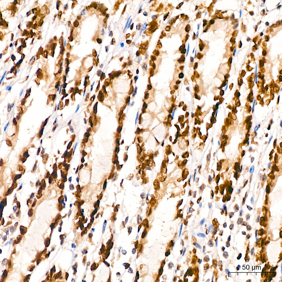
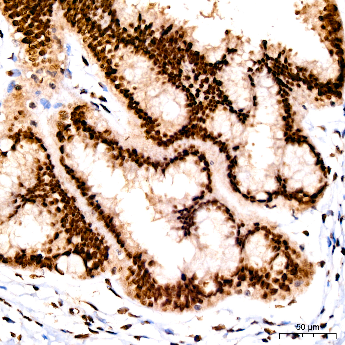
Immunohistochemistry analysis of paraffin-embedded Human cervix using c-Fos Rabbit mAb (A24620) at dilution of 1:400 (40x lens). High pressure antigen retrieval performed with 0.01M Tris/EDTA Buffer (pH 9.0) prior to IHC staining. |
|
|
Immunohistochemistry analysis of paraffin-embedded Human colon using c-Fos Rabbit mAb (A24620) at dilution of 1:400 (40x lens). High pressure antigen retrieval performed with 0.01M Tris/EDTA Buffer (pH 9.0) prior to IHC staining. |
|
|
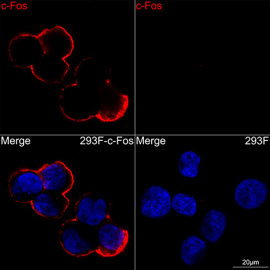
Confocal imaging of paraffin-embedded Mouse brain tissue using c-Fos Rabbit mAb (A24620, dilution 1:200) followed by a further incubation with Cy3 Goat Anti-Rabbit IgG (H+L) (AS007, dilution 1:500) (Red). DAPI was used for nuclear staining (Blue). High pressure antigen retrieval performed with 0.01M Citrate Buffer (pH 6.0) prior to IF staining. Objective: 40x. |
|
|
Confocal imaging of paraffin-embedded Mouse colon tissue using c-Fos Rabbit mAb (A24620, dilution 1:200) followed by a further incubation with Cy3 Goat Anti-Rabbit IgG (H+L) (AS007, dilution 1:500) (Red). DAPI was used for nuclear staining (Blue). High pressure antigen retrieval performed with 0.01M Citrate Buffer (pH 6.0) prior to IF staining. Objective: 40x. |
|
|
Confocal imaging of paraffin-embedded Rat brain tissue using c-Fos Rabbit mAb (A24620, dilution 1:200) followed by a further incubation with Cy3 Goat Anti-Rabbit IgG (H+L) (AS007, dilution 1:500) (Red). DAPI was used for nuclear staining (Blue). High pressure antigen retrieval performed with 0.01M Citrate Buffer (pH 6.0) prior to IF staining. Objective: 40x. |
Product Guarantee and Expert Support
