
PU.1/SPI1 Rabbit mAb, Unconjugated, Monoclonal
Catalog Number:
ABB-A24910
- Images (8)

| Article Name: | PU.1/SPI1 Rabbit mAb, Unconjugated, Monoclonal |
| Biozol Catalog Number: | ABB-A24910 |
| Supplier Catalog Number: | A24910 |
| Alternative Catalog Number: | ABB-A24910-100UL,ABB-A24910-20UL,ABB-A24910-1000UL,ABB-A24910-500UL |
| Manufacturer: | ABclonal |
| Host: | Rabbit |
| Category: | Antikörper |
| Application: | ELISA, FC, IF, IHC-P, WB |
| Species Reactivity: | Human |
| Immunogen: | Synthetic peptide. This information is considered to be commercially sensitive. |
| Conjugation: | Unconjugated |
| Alternative Names: | OF, PU.1, AGM10, SFPI1, SPI-1, SPI-A, PU.1/SPI1 |
| This gene encodes an ETS-domain transcription factor that activates gene expression during myeloid and B-lymphoid cell development. The nuclear protein binds to a purine-rich sequence known as the PU-box found near the promoters of target genes, and regulates their expression in coordination with other transcription factors and cofactors. The protein can also regulate alternative splicing of target genes. Multiple transcript variants encoding different isoforms have been found for this gene. |
| Application Dilute: | WB,1:1000 - 1:6000|IF/ICC,1:50 - 1:200|IF-P,1:50 - 1:200|IHC-P,1:500 - 1:5000|FC (intra),1:100 - 1:500|ELISA,Recommended starting concentration is 1 µg/mL. Please optimize the concentration based on your specific assay requirements. |
| Application Notes: | Cross-Reactivity: Human. ResearchArea: Epigenetics Nuclear Signaling,Transcription Factors,Immunology Inflammation,Cell Intrinsic Innate Immunity Signaling Pathway. Shipping: Ice Bag |
|
|
Immunohistochemistry analysis of paraffin-embedded Human appendix tissue using PU.1/SPI1 Rabbit mAb (A24910) at a dilution of 1:3000 (40x lens). High pressure antigen retrieval performed with 0.01M Tris-EDTA Buffer (pH 9.0) prior to IHC staining. |
|
|
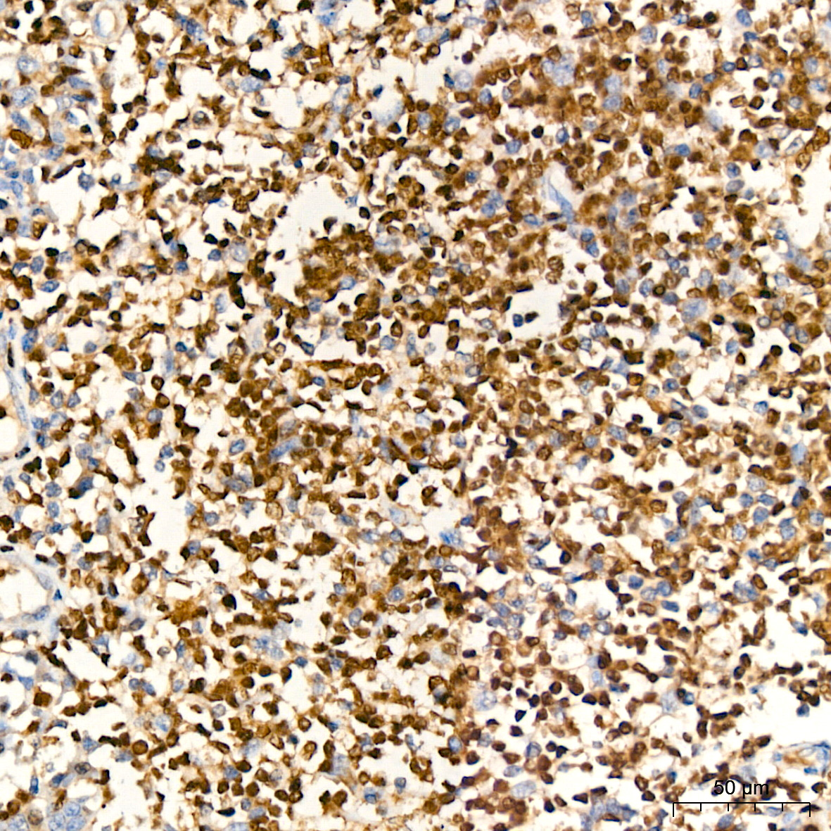
Immunohistochemistry analysis of paraffin-embedded Human follicular lymphoma tissue using PU.1/SPI1 Rabbit mAb (A24910) at a dilution of 1:3000 (40x lens). High pressure antigen retrieval performed with 0.01M Tris-EDTA Buffer (pH 9.0) prior to IHC staining. |
|
|
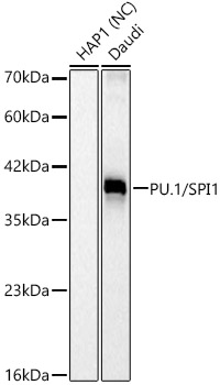
Western blot analysis of various lysates, using PU.1/SPI1 Rabbit mAb (A24910) at 1:1000 dilution. Secondary antibody: HRP-conjugated Goat anti-Rabbit IgG (H+L) (AS014) at 1:10000 dilution. Lysates/proteins: 25µg per lane. Blocking buffer: 3% nonfat dry milk in TBST. Detection: ECL Basic Kit (RM00020). Negative control (NC):HAP1 Exposure time: 15s. |
|
|
Immunohistochemistry analysis of paraffin-embedded Human tonsil tissue using PU.1/SPI1 Rabbit mAb (A24910) at a dilution of 1:3000 (40x lens). High pressure antigen retrieval performed with 0.01M Tris-EDTA Buffer (pH 9.0) prior to IHC staining. |
|
|
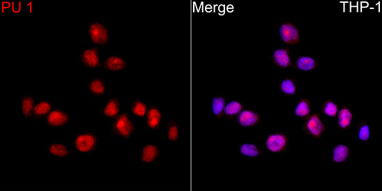
Confocal imaging of THP-1 cells using PU.1/SPI1 Rabbit mAb (A24910, dilution 1:200) followed by a further incubation with Cy3 Goat Anti-Rabbit IgG (H+L) (AS007, dilution 1:500) (Red). DAPI was used for nuclear staining (Blue). Objective: 100x. |
|
|
Confocal imaging of paraffin-embedded Human spleen tissue using PU.1/SPI1 Rabbit mAb (A24910, dilution 1:200) followed by a further incubation with Cy3 Goat Anti-Rabbit IgG (H+L) (AS007, dilution 1:500) (Red). DAPI was used for nuclear staining (Blue). High pressure antigen retrieval performed with 0.01M Citrate Buffer (pH 6.0) prior to IF staining. Objective: 40x. |
|
|
Flow cytometry: 1X10 6 HAP1 cells (negative control,left)and THP-1 cells (right) were intracellularly-stained with PU.1/SPI1 Rabbit mAb (A24910,2 µg/mL,orange line) or ABflo 594 Rabbit IgG isotype control (A23821,5 µl/Test,blue line), followed by ABflo 594-conjugated Goat Anti-Rabbit IgG (H+L) staining. Non-fluorescently stained cells were used as blank control (red line). |
|
|
Flow cytometry: 1X10 6 THP-1 cells were intracellularly-stained with ABflo 594 Rabbit IgG isotype control (A23821,5 µl/Test,left) or PU.1/SPI1 Rabbit mAb (A24910,2 µg/mL,right). |
Product Guarantee and Expert Support
