
N-WASP/WASL Rabbit pAb, Unconjugated, Polyclonal
Catalog Number:
ABB-A2576
- Images (8)


| Article Name: | N-WASP/WASL Rabbit pAb, Unconjugated, Polyclonal |
| Biozol Catalog Number: | ABB-A2576 |
| Supplier Catalog Number: | A2576 |
| Alternative Catalog Number: | ABB-A2576-100UL,ABB-A2576-500UL,ABB-A2576-1000UL,ABB-A2576-20UL |
| Manufacturer: | ABclonal |
| Host: | Rabbit |
| Category: | Antikörper |
| Application: | ELISA, IF, IHC-P, WB |
| Species Reactivity: | Human |
| Immunogen: | Recombinant protein (or fragment).This information is considered to be commercially sensitive. |
| Conjugation: | Unconjugated |
| Alternative Names: | NWASP, WASPB, N-WASP, N-WASP/WASL |
| This gene encodes a member of the Wiskott-Aldrich syndrome (WAS) protein family. Wiskott-Aldrich syndrome proteins share similar domain structure, and associate with a variety of signaling molecules to alter the actin cytoskeleton. The encoded protein is highly expressed in neural tissues, and interacts with several proteins involved in cytoskeletal organization, including cell division control protein 42 (CDC42) and the actin-related protein-2/3 (ARP2/3) complex. The encoded protein may be involved in the formation of long actin microspikes, and in neurite extension. |
| Clonality: | Polyclonal |
| Molecular Weight: | 55kDa |
| NCBI: | 8976 |
| UniProt: | O00401 |
| Purity: | Affinity purification |
| Sequence: | MSSVQQQPPPPRRVTNVGSLLLTPQENESLFTFLGKKCVTMSSAVVQLYAADRNCMWSKKCSGVACLVKDNPQRSYFLRIFDIKDGKLLWEQELYNNFVYNSPRGYFHTFAGDTCQVALNFANEEEAKKFRKAVTDLLGRRQRKSEKRRDPPNGPNLPMATVDIKNPEITTNRFYGPQVNNISHTKEKKKGKAKKKRLTKADIGTPSNFQHIGHVGWDPNTGFDLNNLDPELKNLFDMCGISEAQLKDRE |
| Target: | WASL |
| Antibody Type: | Primary Antibody |
| Application Dilute: | WB,1:500 - 1:2000|IHC-P,1:50 - 1:200|IF/ICC,1:50 - 1:200|ELISA,Recommended starting concentration is 1 µg/mL. Please optimize the concentration based on your specific assay requirements. |
| Application Notes: | Cross-Reactivity: Human,Mouse,Rat. ResearchArea: Epigenetics Nuclear Signaling,Signal Transduction,Cell Biology Developmental Biology,Cytoskeleton,Microfilaments. Shipping: Ice Bag |
|
|
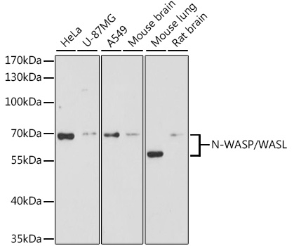
Western blot analysis of lysates from HeLa cells using N-WASP/WASL Rabbit pAb (A2576) at 1:3000 dilution incubated overnight at 4°C. Secondary antibody: HRP-conjugated Goat anti-Rabbit IgG (H+L) (AS014) at 1:10000 dilution. Lysates/proteins: 25 µg per lane. Blocking buffer: 3% nonfat dry milk in TBST. Detection: ECL Basic Kit (RM00020). Exposure time: 30s. |
|
|
Western blot analysis of lysates from Mouse lung using N-WASP/WASL Rabbit pAb (A2576) at 1:3000 dilution incubated overnight at 4°C. Secondary antibody: HRP-conjugated Goat anti-Rabbit IgG (H+L) (AS014) at 1:10000 dilution. Lysates/proteins: 25 µg per lane. Blocking buffer: 3% nonfat dry milk in TBST. Detection: ECL Basic Kit (RM00020). Exposure time: 90s. |
|
|
Western blot analysis of lysates from Rat kidney usingN-WASP/WASL Rabbit pAb (A2576) at1:2000 dilution. Secondary antibody: HRP-conjugated Goat anti-Rabbit IgG (H+L) (AS014) at 1:10000 dilution. Lysates/proteins: 25µg per lane. Blocking buffer: 3% nonfat dry milk in TBST. Detection: ECL Basic Kit (RM00020). Exposure time: 30s. |
|
|
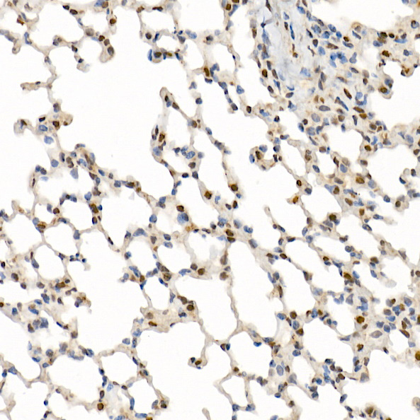
Immunohistochemistry analysis of paraffin-embedded Mouse lung using N-WASP/WASL Rabbit pAb (A2576) at dilution of 1:200 (40x lens). High pressure antigen retrieval performed with 0.01M Citrate buffer (pH 6.0) prior to IHC staining. |
|
|
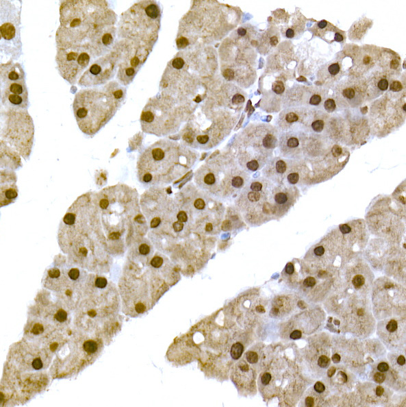
Immunohistochemistry analysis of paraffin-embedded Mouse pancreas using N-WASP/WASL Rabbit pAb (A2576) at dilution of 1:200 (40x lens). High pressure antigen retrieval performed with 0.01M Citrate buffer (pH 6.0) prior to IHC staining. |
|
|
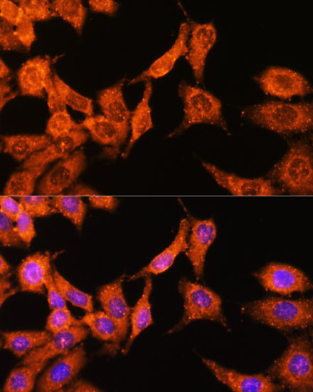
Immunofluorescence analysis of C6 cells using N-WASP/WASL Rabbit pAb (A2576) at dilution of 1:100 (40x lens). Secondary antibody: Cy3-conjugated Goat anti-Rabbit IgG (H+L) (AS007) at 1:500 dilution. Blue: DAPI for nuclear staining. |
|
|
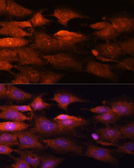
Immunofluorescence analysis of HeLa cells using N-WASP/WASL Rabbit pAb (A2576) at dilution of 1:100 (40x lens). Secondary antibody: Cy3-conjugated Goat anti-Rabbit IgG (H+L) (AS007) at 1:500 dilution. Blue: DAPI for nuclear staining. |
|
|
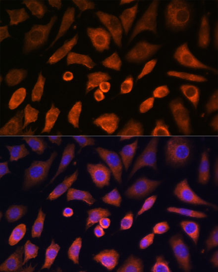
Immunofluorescence analysis of L-929 cells using N-WASP/WASL Rabbit pAb (A2576) at dilution of 1:100 (40x lens). Secondary antibody: Cy3-conjugated Goat anti-Rabbit IgG (H+L) (AS007) at 1:500 dilution. Blue: DAPI for nuclear staining. |
Product Guarantee and Expert Support
