

[KO Validated] TBK1/NAK Rabbit mAb, Unconjugated, Monoclonal
Catalog Number:
ABB-A3458
- Images (9)

| Article Name: | [KO Validated] TBK1/NAK Rabbit mAb, Unconjugated, Monoclonal |
| Biozol Catalog Number: | ABB-A3458 |
| Supplier Catalog Number: | A3458 |
| Alternative Catalog Number: | ABB-A3458-20UL,ABB-A3458-100UL,ABB-A3458-1000UL,ABB-A3458-500UL |
| Manufacturer: | ABclonal |
| Host: | Rabbit |
| Category: | Antikörper |
| Application: | ELISA, IF, IHC-P, IP, WB |
| Species Reactivity: | Human |
| Immunogen: | Synthetic peptide. This information is considered to be commercially sensitive. |
| Conjugation: | Unconjugated |
| Alternative Names: | NAK, T2K, IIAE8, FTDALS4, TBK1/NAK |
| The NF-kappa-B (NFKB) complex of proteins is inhibited by I-kappa-B (IKB) proteins, which inactivate NFKB by trapping it in the cytoplasm. Phosphorylation of serine residues on the IKB proteins by IKB kinases marks them for destruction via the ubiquitination pathway, thereby allowing activation and nuclear translocation of the NFKB complex. The protein encoded by this gene is similar to IKB kinases and can mediate NFKB activation in response to certain growth factors. The protein is also an important kinase for antiviral innate immunity response. |
| Application Dilute: | WB,1:4000 - 1:15000|IF/ICC,1:100 - 1:800|IHC-P,1:500 - 1:1000|IP,0.5µg-4µg antibody for 200µg-400µg extracts of whole cells|ELISA,Recommended starting concentration is 1 µg/mL. Please optimize the concentration based on your specific assay requirements. |
| Application Notes: | Cross-Reactivity: Human,Mouse,Rat. ResearchArea: Epigenetics Nuclear Signaling,Protein phosphorylation,Signal Transduction,Kinase,Serine threonine kinases,PI3K-Akt Signaling Pathway,Immunology Inflammation,NF-kB Signaling Pathway,Toll-like Receptor Signaling Pathway. Shipping: Ice Bag |
|
|
Western blot analysis of lysates from wild type (WT) and TBK1/NAK knockout (KO) 293T cells using [KO Validated] TBK1/NAK Rabbit mAb (A3458) at 1:7000 dilution incubated at room temperature for 1.5 hours. Secondary antibody: HRP-conjugated Goat anti-Rabbit IgG (H+L) (AS014) at 1:10000 dilution. Lysates/proteins: 25 µg per lane. Blocking buffer: 3% nonfat dry milk in TBST. Detection: ECL Basic Kit (RM00020). Exposure time: 90 s. |
|
|
Western blot analysis of lysates from Raji cells using [KO Validated] TBK1/NAK Rabbit mAb (A3458) at 1:7000 dilution incubated at room temperature for 1.5 hours. Secondary antibody: HRP-conjugated Goat anti-Rabbit IgG (H+L) (AS014) at 1:10000 dilution. Lysates/proteins: 25 µg per lane. Blocking buffer: 3% nonfat dry milk in TBST. Detection: ECL Basic Kit (RM00020). Exposure time: 90 s. |
|
|
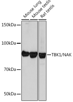
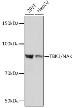
Western blot analysis of various lysates using [KO Validated] TBK1/NAK Rabbit mAb (A3458) at 1:4000 dilution incubated at room temperature for 1.5 hours. Secondary antibody: HRP-conjugated Goat anti-Rabbit IgG (H+L) (AS014) at 1:10000 dilution. Lysates/proteins: 25 µg per lane. Blocking buffer: 3% nonfat dry milk in TBST. Detection: ECL Basic Kit (RM00020). Exposure time: 90 s. |
|
|
Immunohistochemistry analysis of paraffin-embedded Rat testis tissue using [KO Validated] TBK1/NAK Rabbit mAb (A3458) at a dilution of 1:750 (40x lens). High pressure antigen retrieval performed with 0.01M Tris-EDTA Buffer (pH 9.0) prior to IHC staining. |
|
|
Immunohistochemistry analysis of paraffin-embedded Human testis tissue using [KO Validated] TBK1/NAK Rabbit mAb (A3458) at a dilution of 1:750 (40x lens). High pressure antigen retrieval performed with 0.01M Tris-EDTA Buffer (pH 9.0) prior to IHC staining. |
|
|
Immunohistochemistry analysis of paraffin-embedded Mouse testis tissue using [KO Validated] TBK1/NAK Rabbit mAb (A3458) at a dilution of 1:750 (40x lens). High pressure antigen retrieval performed with 0.01M Tris-EDTA Buffer (pH 9.0) prior to IHC staining. |
|
|
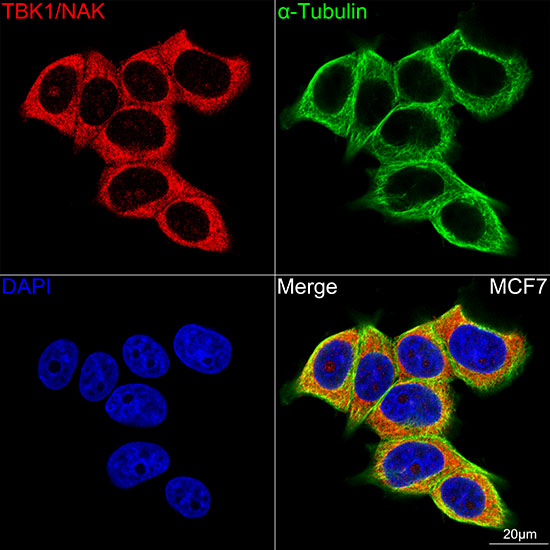
Confocal imaging of MCF7 cells using [KO Validated] TBK1/NAK Rabbit mAb (A3458,at dilution of 1:100) (Red). The cells were counterstained with alpha-Tubulin Mouse mAb (AC012,dilution 1:400) (Green). DAPI was used for nuclear staining (blue). Objective: 100x. |
|
|
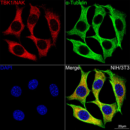
Confocal imaging of NIH/3T3 cells using [KO Validated] TBK1/NAK Rabbit mAb (A3458,at dilution of 1:100) (Red). The cells were counterstained with alpha-Tubulin Mouse mAb (AC012,dilution 1:400) (Green). DAPI was used for nuclear staining (blue). Objective: 100x. |
|
|
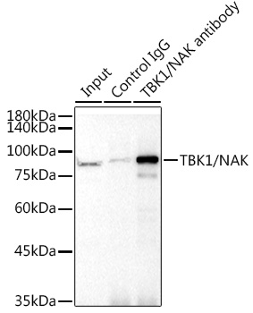
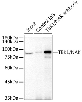
Immunoprecipitation analysis of 300 µg extracts from 293T cells using 3 µg [KO Validated] TBK1/NAK Rabbit mAb (A3458). Western blot was performed from the immunoprecipitate using [KO Validated] TBK1/NAK Rabbit mAb (A3458) at a dilution of 1:1000. |
Product Guarantee and Expert Support
