
LXRalpha Rabbit mAb, Unconjugated, Monoclonal
Catalog Number:
ABB-A3974
- Images (8)

| Article Name: | LXRalpha Rabbit mAb, Unconjugated, Monoclonal |
| Biozol Catalog Number: | ABB-A3974 |
| Supplier Catalog Number: | A3974 |
| Alternative Catalog Number: | ABB-A3974-20UL,ABB-A3974-100UL,ABB-A3974-500UL,ABB-A3974-1000UL |
| Manufacturer: | ABclonal |
| Host: | Rabbit |
| Category: | Antikörper |
| Application: | ELISA, IF, IHC-P, WB |
| Species Reactivity: | Human |
| Immunogen: | Synthetic peptide. This information is considered to be commercially sensitive. |
| Conjugation: | Unconjugated |
| Alternative Names: | LXRA, LXR-a, RLD-1, LXRalpha |
| The protein encoded by this gene belongs to the NR1 subfamily of the nuclear receptor superfamily. The NR1 family members are key regulators of macrophage function, controlling transcriptional programs involved in lipid homeostasis and inflammation. This protein is highly expressed in visceral organs, including liver, kidney and intestine. It forms a heterodimer with retinoid X receptor (RXR), and regulates expression of target genes containing retinoid response elements. Studies in mice lacking this gene suggest that it may play an important role in the regulation of cholesterol homeostasis. Alternatively spliced transcript variants encoding different isoforms have been found for this gene. |
| Application Dilute: | WB,1:1000 - 1:6000|IHC-P,1:100 - 1:1000|IF/ICC,1:50 - 1:200|ELISA,Recommended starting concentration is 1 µg/mL. Please optimize the concentration based on your specific assay requirements. |
| Application Notes: | Cross-Reactivity: Human,Mouse,Rat. ResearchArea: Epigenetics Nuclear Signaling,Transcription Factors,Nuclear Receptor Signaling,Cancer,Signal Transduction,Endocrine Metabolism,Lipid Metabolism,Cholesterol Metabolism,Cardiovascular,Lipids. Shipping: Ice Bag |
|
|
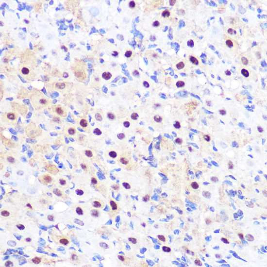
Immunohistochemistry analysis of paraffin-embedded Rat ovary using LXRalpha Rabbit mAb (A3974) at dilution of 1:100 (40x lens). Microwave antigen retrieval performed with 0.01M PBS Buffer (pH 7.2) prior to IHC staining. |
|
|
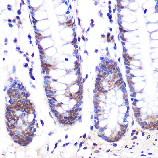
Immunohistochemistry analysis of paraffin-embedded Human colon using LXRalpha Rabbit mAb (A3974) at dilution of 1:100 (40x lens). Microwave antigen retrieval performed with 0.01M PBS Buffer (pH 7.2) prior to IHC staining. |
|
|
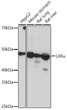
Western blot analysis of various lysates using LXRalpha Rabbit mAb (A3974) at 1:1000 dilution. Secondary antibody: HRP-conjugated Goat anti-Rabbit IgG (H+L) (AS014) at 1:10000 dilution. Lysates/proteins: 25µg per lane. Blocking buffer: 3% nonfat dry milk in TBST. Detection: ECL Basic Kit (RM00020). Exposure time: 3s. |
|
|
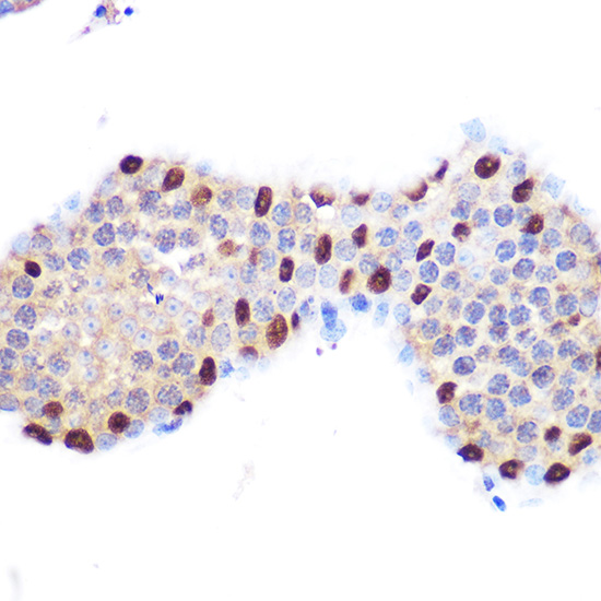
Immunohistochemistry analysis of paraffin-embedded Mouse testis using LXRalpha Rabbit mAb (A3974) at dilution of 1:100 (40x lens). Microwave antigen retrieval performed with 0.01M PBS Buffer (pH 7.2) prior to IHC staining. |
|
|
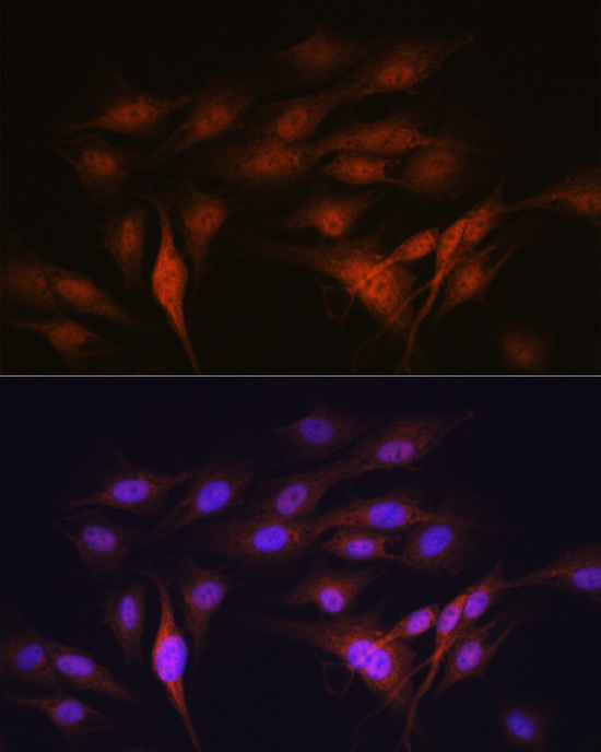
Immunofluorescence analysis of C6 cells using LXRalpha Rabbit mAb (A3974) at dilution of 1:100 (40x lens). Secondary antibody: Cy3-conjugated Goat anti-Rabbit IgG (H+L) (AS007) at 1:500 dilution. Blue: DAPI for nuclear staining. |
|
|
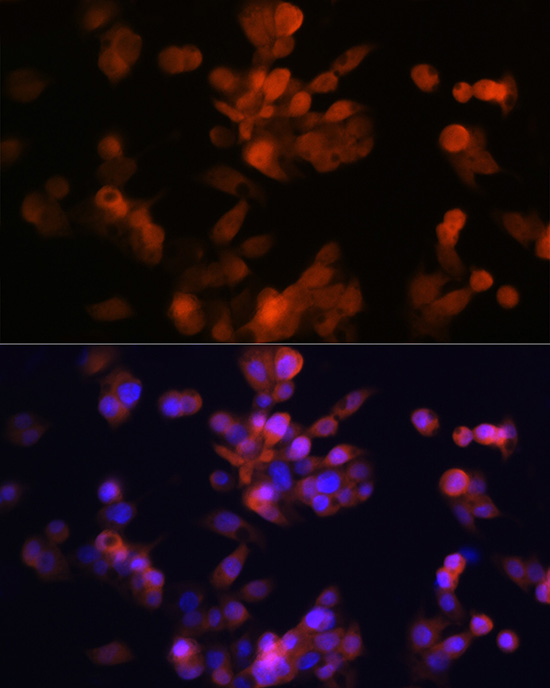
Immunofluorescence analysis of Hep G2 cells using LXRalpha Rabbit mAb (A3974) at dilution of 1:100 (40x lens). Secondary antibody: Cy3-conjugated Goat anti-Rabbit IgG (H+L) (AS007) at 1:500 dilution. Blue: DAPI for nuclear staining. |
|
|
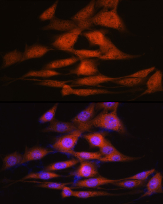
Immunofluorescence analysis of NIH-3T3 cells using LXRalpha Rabbit mAb (A3974) at dilution of 1:100 (40x lens). Secondary antibody: Cy3-conjugated Goat anti-Rabbit IgG (H+L) (AS007) at 1:500 dilution. Blue: DAPI for nuclear staining. |
|
|
Confocal imaging of C2C12 cells usingLXRalpha Rabbit mAb (A3974, dilution 1:100) followed by a further incubation with Cy3 Goat Anti-Rabbit IgG (H+L) (AS007, dilution 1:500) (Red). The cells were counterstained with alpha-Tubulin Mouse mAb (AC012, dilution 1:400) followed by incubation with ABflo 488-conjugated Goat Anti-Mouse IgG (H+L) Ab (AS076, dilution 1:500) (Green). DAPI was used for nuclear staining (Blue). Objective: 100x. |
Product Guarantee and Expert Support
