
UBE2I Rabbit mAb, Unconjugated, Monoclonal
Catalog Number:
ABB-A4396
- Images (9)

| Article Name: | UBE2I Rabbit mAb, Unconjugated, Monoclonal |
| Biozol Catalog Number: | ABB-A4396 |
| Supplier Catalog Number: | A4396 |
| Alternative Catalog Number: | ABB-A4396-100UL,ABB-A4396-20UL |
| Manufacturer: | ABclonal |
| Host: | Rabbit |
| Category: | Antikörper |
| Application: | ELISA, IHC-P, WB |
| Species Reactivity: | Human |
| Immunogen: | Synthetic peptide. This information is considered to be commercially sensitive. |
| Conjugation: | Unconjugated |
| Alternative Names: | P18, UBC9, C358B7.1, UBE2I |
| The modification of proteins with ubiquitin is an important cellular mechanism for targeting abnormal or short-lived proteins for degradation. Ubiquitination involves at least three classes of enzymes: ubiquitin-activating enzymes, or E1s, ubiquitin-conjugating enzymes, or E2s, and ubiquitin-protein ligases, or E3s. This gene encodes a member of the E2 ubiquitin-conjugating enzyme family. Four alternatively spliced transcript variants encoding the same protein have been found for this gene. |
| Application Dilute: | WB,1:500 - 1:1000|IHC-P,1:50 - 1:200|ELISA,Recommended starting concentration is 1 µg/mL. Please optimize the concentration based on your specific assay requirements. |
| Application Notes: | Cross-Reactivity: Human,Mouse,Rat. ResearchArea: Epigenetics Nuclear Signaling,RNA Binding,Cell Biology Developmental Biology,Apoptosis,Ubiquitin,Ubiquitin-Proteasome Signaling Pathway,Endocrine Metabolism,Endocrine and metabolic diseases,Obesity. Shipping: Ice Bag |
|
|
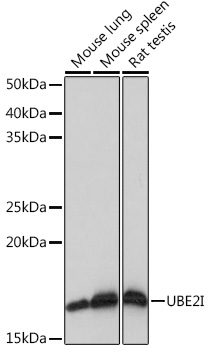
Western blot analysis of various lysates using UBE2I Rabbit mAb (A4396) at 1:1000 dilution. Secondary antibody: HRP-conjugated Goat anti-Rabbit IgG (H+L) (AS014) at 1:10000 dilution. Lysates/proteins: 25µg per lane. Blocking buffer: 3% nonfat dry milk in TBST. Detection: ECL Basic Kit (RM00020). Exposure time: 1s. |
|
|
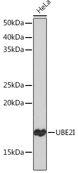
Western blot analysis of lysates from HeLa cells, using UBE2I Rabbit mAb (A4396) at 1:1000 dilution. Secondary antibody: HRP-conjugated Goat anti-Rabbit IgG (H+L) (AS014) at 1:10000 dilution. Lysates/proteins: 25µg per lane. Blocking buffer: 3% nonfat dry milk in TBST. Detection: ECL Basic Kit (RM00020). Exposure time: 30s. |
|
|
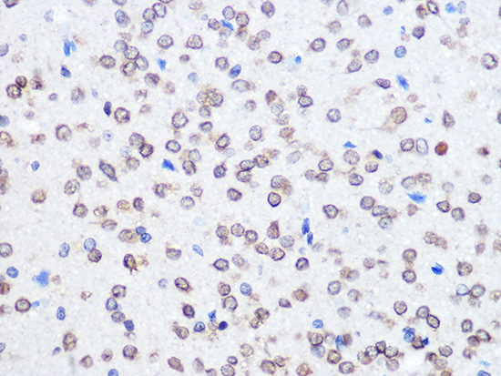
Immunohistochemistry analysis of paraffin-embedded Human thyroid cancer tissue using UBE2I Rabbit mAb (A4396) at a dilution of 1:200 (40x lens). High pressure antigen retrieval performed with 0.01M Citrate buffer (pH 6.0) prior to IHC staining. |
|
|
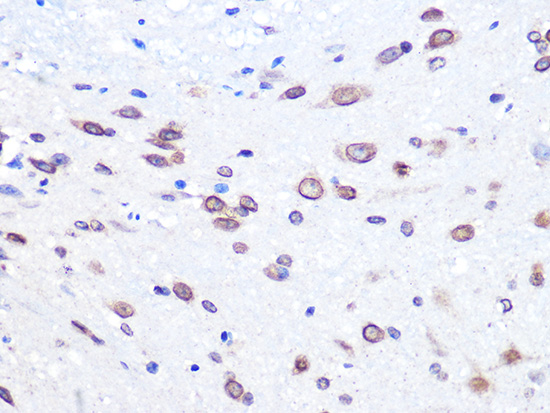
Immunohistochemistry analysis of paraffin-embedded Mouse brain tissue using UBE2I Rabbit mAb (A4396) at a dilution of 1:200 (40x lens). High pressure antigen retrieval performed with 0.01M Citrate buffer (pH 6.0) prior to IHC staining. |
|
|
Immunohistochemistry analysis of paraffin-embedded Mouse lung tissue using UBE2I Rabbit mAb (A4396) at a dilution of 1:200 (40x lens). High pressure antigen retrieval performed with 0.01M Citrate buffer (pH 6.0) prior to IHC staining. |
|
|
Immunohistochemistry analysis of paraffin-embedded Mouse spleen tissue using UBE2I Rabbit mAb (A4396) at a dilution of 1:200 (40x lens). High pressure antigen retrieval performed with 0.01M Citrate buffer (pH 6.0) prior to IHC staining. |
|
|
Immunohistochemistry analysis of paraffin-embedded Mouse testis tissue using UBE2I Rabbit mAb (A4396) at a dilution of 1:200 (40x lens). High pressure antigen retrieval performed with 0.01M Citrate buffer (pH 6.0) prior to IHC staining. |
|
|
Immunohistochemistry analysis of paraffin-embedded Rat brain tissue using UBE2I Rabbit mAb (A4396) at a dilution of 1:200 (40x lens). High pressure antigen retrieval performed with 0.01M Citrate buffer (pH 6.0) prior to IHC staining. |
|
|
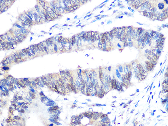
Immunohistochemistry analysis of paraffin-embedded Rat colon tissue using UBE2I Rabbit mAb (A4396) at a dilution of 1:200 (40x lens). High pressure antigen retrieval performed with 0.01M Citrate buffer (pH 6.0) prior to IHC staining. |
Product Guarantee and Expert Support
