
PBR/TSPO Rabbit mAb, Unconjugated, Monoclonal
Catalog Number:
ABB-A4881
- Images (9)

| Article Name: | PBR/TSPO Rabbit mAb, Unconjugated, Monoclonal |
| Biozol Catalog Number: | ABB-A4881 |
| Supplier Catalog Number: | A4881 |
| Alternative Catalog Number: | ABB-A4881-100UL,ABB-A4881-20UL |
| Manufacturer: | ABclonal |
| Host: | Rabbit |
| Category: | Antikörper |
| Application: | ELISA, IF, IHC-P, WB |
| Species Reactivity: | Human |
| Immunogen: | Synthetic peptide. This information is considered to be commercially sensitive. |
| Conjugation: | Unconjugated |
| Alternative Names: | DBI, IBP, MBR, PBR, PBS, BPBS, BZRP, PKBS, PTBR, mDRC, pk18, TSPO1, PBR/TSPO |
| Present mainly in the mitochondrial compartment of peripheral tissues, the protein encoded by this gene interacts with some benzodiazepines and has different affinities than its endogenous counterpart. The protein is a key factor in the flow of cholesterol into mitochondria to permit the initiation of steroid hormone synthesis. Alternatively spliced transcript variants have been reported, one of the variants lacks an internal exon and is considered non-coding, and the other variants encode the same protein. |
| Application Dilute: | WB,1:5000 - 1:30000|IHC-P,1:200 - 1:2000|IF/ICC,1:100 - 1:400|ELISA,Recommended starting concentration is 1 µg/mL. Please optimize the concentration based on your specific assay requirements. |
| Application Notes: | Cross-Reactivity: Human,Mouse. ResearchArea: Cancer,Signal Transduction,Cell Biology Developmental Biology,Apoptosis,Endocrine Metabolism,Mitochondrial metabolism,Mitochondrial markers,Lipid Metabolism,Cholesterol Metabolism,Neuroscience,Calcium Signaling,Cardiovascular,Lipids. Shipping: Ice Bag |
|
|
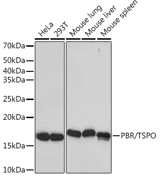
Western blot analysis of various lysates using PBR/TSPO Rabbit mAb (A4881) at 1:5000 dilution incubated overnight at 4°C. Secondary antibody: HRP-conjugated Goat anti-Rabbit IgG (H+L) (AS014) at 1:10000 dilution. Lysates/proteins: 25 µg per lane. Blocking buffer: 3% nonfat dry milk in TBST. Detection: ECL Basic Kit (RM00020). Exposure time: 1s. |
|
|
Immunohistochemistry analysis of paraffin-embedded Human kidney tissue using PBR/TSPO Rabbit mAb (A4881) at a dilution of 1:200 (40x lens). High pressure antigen retrieval performed with 0.01M Citrate Buffer (pH 6.0) prior to IHC staining. |
|
|
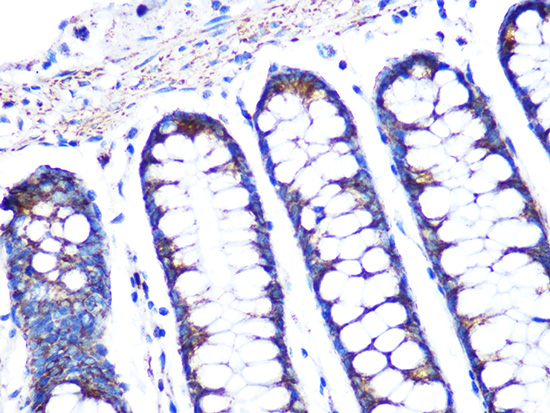
Immunohistochemistry analysis of paraffin-embedded Human colon carcinoma tissue using PBR/TSPO Rabbit mAb (A4881) at a dilution of 1:200 (40x lens). High pressure antigen retrieval performed with 0.01M Citrate Buffer (pH 6.0) prior to IHC staining. |
|
|
Immunohistochemistry analysis of paraffin-embedded Human liver tissue using PBR/TSPO Rabbit mAb (A4881) at a dilution of 1:200 (40x lens). High pressure antigen retrieval performed with 0.01M Citrate Buffer (pH 6.0) prior to IHC staining. |
|
|
Immunohistochemistry analysis of paraffin-embedded Human lung cancer tissue using PBR/TSPO Rabbit mAb (A4881) at a dilution of 1:200 (40x lens). High pressure antigen retrieval performed with 0.01M Citrate Buffer (pH 6.0) prior to IHC staining. |
|
|
Immunohistochemistry analysis of paraffin-embedded Mouse kidney tissue using PBR/TSPO Rabbit mAb (A4881) at a dilution of 1:200 (40x lens). High pressure antigen retrieval performed with 0.01M Citrate Buffer (pH 6.0) prior to IHC staining. |
|
|
Immunohistochemistry analysis of paraffin-embedded Mouse liver tissue using PBR/TSPO Rabbit mAb (A4881) at a dilution of 1:200 (40x lens). High pressure antigen retrieval performed with 0.01M Citrate Buffer (pH 6.0) prior to IHC staining. |
|
|
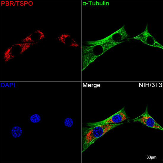
Confocal imaging of NIH/3T3 cells using PBR/TSPO Rabbit mAb (A4881,dilution 1:100)(Red). The cells were counterstained with alpha-Tubulin Mouse mAb (AC012,dilution 1:400) (Green). DAPI was used for nuclear staining (blue). Objective: 100x. |
|
|
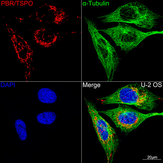
Confocal imaging of U-2 OS cells using PBR/TSPO Rabbit mAb (A4881,dilution 1:100)(Red). The cells were counterstained with alpha-Tubulin Mouse mAb (AC012,dilution 1:400) (Green). DAPI was used for nuclear staining (blue). Objective: 100x. |
Product Guarantee and Expert Support
