
HSPE1/HSP10/CPN10 Rabbit mAb, Unconjugated, Monoclonal
Catalog Number:
ABB-A5580
- Images (9)


| Article Name: | HSPE1/HSP10/CPN10 Rabbit mAb, Unconjugated, Monoclonal |
| Biozol Catalog Number: | ABB-A5580 |
| Supplier Catalog Number: | A5580 |
| Alternative Catalog Number: | ABB-A5580-100UL,ABB-A5580-20UL |
| Manufacturer: | ABclonal |
| Host: | Rabbit |
| Category: | Antikörper |
| Application: | ELISA, IHC-P, WB |
| Species Reactivity: | Human |
| Immunogen: | Recombinant protein (or fragment).This information is considered to be commercially sensitive. |
| Conjugation: | Unconjugated |
| Alternative Names: | EPF, CPN10, GROES, HSP10, HSPE1/HSP10/CPN10 |
| This gene encodes a major heat shock protein which functions as a chaperonin. Its structure consists of a heptameric ring which binds to another heat shock protein in order to form a symmetric, functional heterodimer which enhances protein folding in an ATP-dependent manner. This gene and its co-chaperonin, HSPD1, are arranged in a head-to-head orientation on chromosome 2. Naturally occurring read-through transcription occurs between this locus and the neighboring locus MOBKL3. |
| Application Dilute: | WB,1:1000 - 1:6000|IHC-P,1:100 - 1:500|ELISA,Recommended starting concentration is 1 µg/mL. Please optimize the concentration based on your specific assay requirements. |
| Application Notes: | Cross-Reactivity: Human,Mouse,Rat. ResearchArea: Epigenetics Nuclear Signaling,RNA Binding,Signal Transduction,Cell Biology Developmental Biology,Apoptosis,Endocrine Metabolism,Mitochondrial metabolism,Mitochondrial markers. Shipping: Ice Bag |
|
|
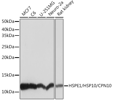
Western blot analysis of various lysates using HSPE1/HSP10/HSPE1/HSP10/CPN10 Rabbit mAb (A5580) at 1:1000 dilution. Secondary antibody: HRP-conjugated Goat anti-Rabbit IgG (H+L) (AS014) at 1:10000 dilution. Lysates/proteins: 25µg per lane. Blocking buffer: 3% nonfat dry milk in TBST. Detection: ECL Basic Kit (RM00020). Exposure time: 1s. |
|
|
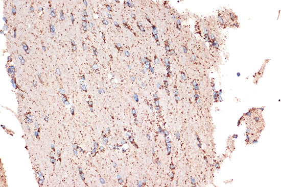
Immunohistochemistry analysis of paraffin-embedded Rat brain using HSPE1/HSP10/HSPE1/HSP10/CPN10 Rabbit mAb (A5580) at dilution of 1:100 (40x lens). Microwave antigen retrieval performed with 0.01M Tris/EDTA Buffer (pH 9.0) prior to IHC staining. |
|
|
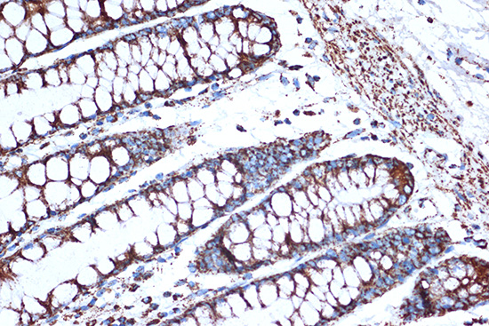
Immunohistochemistry analysis of paraffin-embedded Human colon using HSPE1/HSP10/HSPE1/HSP10/CPN10 Rabbit mAb (A5580) at dilution of 1:100 (40x lens). Microwave antigen retrieval performed with 0.01M Tris/EDTA Buffer (pH 9.0) prior to IHC staining. |
|
|
Immunohistochemistry analysis of paraffin-embedded Human placenta using HSPE1/HSP10/HSPE1/HSP10/CPN10 Rabbit mAb (A5580) at dilution of 1:100 (40x lens). Microwave antigen retrieval performed with 0.01M Tris/EDTA Buffer (pH 9.0) prior to IHC staining. |
|
|
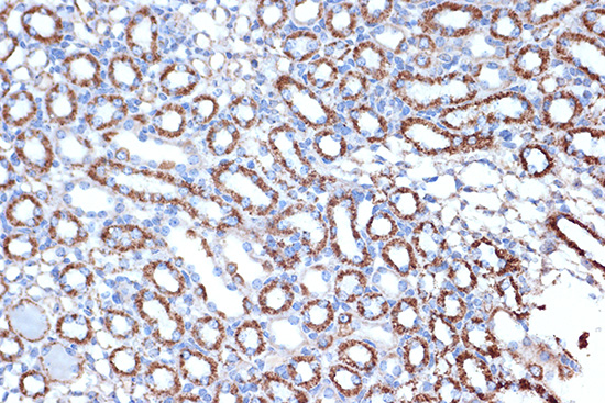
Immunohistochemistry analysis of paraffin-embedded Mouse kidney using HSPE1/HSP10/HSPE1/HSP10/CPN10 Rabbit mAb (A5580) at dilution of 1:100 (40x lens). Microwave antigen retrieval performed with 0.01M Tris/EDTA Buffer (pH 9.0) prior to IHC staining. |
|
|
Immunohistochemistry analysis of paraffin-embedded Human thyroid cancer tissue using HSPE1/HSP10/CPN10 Rabbit mAb (A5580) at a dilution of 1:400 (40x lens). High pressure antigen retrieval performed with 0.01M Citrate buffer (pH 6.0) prior to IHC staining. |
|
|
Immunohistochemistry analysis of paraffin-embedded Human liver tissue using HSPE1/HSP10/CPN10 Rabbit mAb (A5580) at a dilution of 1:400 (40x lens). High pressure antigen retrieval performed with 0.01M Citrate buffer (pH 6.0) prior to IHC staining. |
|
|
Immunohistochemistry analysis of paraffin-embedded Mouse colon tissue using HSPE1/HSP10/CPN10 Rabbit mAb (A5580) at a dilution of 1:400 (40x lens). High pressure antigen retrieval performed with 0.01M Citrate buffer (pH 6.0) prior to IHC staining. |
|
|
Immunohistochemistry analysis of paraffin-embedded Rat kidney tissue using HSPE1/HSP10/CPN10 Rabbit mAb (A5580) at a dilution of 1:400 (40x lens). High pressure antigen retrieval performed with 0.01M Citrate buffer (pH 6.0) prior to IHC staining. |
Product Guarantee and Expert Support
