
[KD Validated] Smad4 Rabbit pAb, Unconjugated, Polyclonal
Catalog Number:
ABB-A5657
- Images (9)

| Article Name: | [KD Validated] Smad4 Rabbit pAb, Unconjugated, Polyclonal |
| Biozol Catalog Number: | ABB-A5657 |
| Supplier Catalog Number: | A5657 |
| Alternative Catalog Number: | ABB-A5657-500UL,ABB-A5657-100UL,ABB-A5657-20UL,ABB-A5657-1000UL |
| Manufacturer: | ABclonal |
| Host: | Rabbit |
| Category: | Antikörper |
| Application: | ELISA, IF, IHC-P, WB |
| Species Reactivity: | Human |
| Immunogen: | Recombinant protein (or fragment).This information is considered to be commercially sensitive. |
| Conjugation: | Unconjugated |
| Alternative Names: | JIP, DPC4, MADH4, MYHRS, Smad4 |
| This gene encodes a member of the Smad family of signal transduction proteins. Smad proteins are phosphorylated and activated by transmembrane serine-threonine receptor kinases in response to transforming growth factor (TGF)-beta signaling. The product of this gene forms homomeric complexes and heteromeric complexes with other activated Smad proteins, which then accumulate in the nucleus and regulate the transcription of target genes. This protein binds to DNA and recognizes an 8-bp palindromic sequence (GTCTAGAC) called the Smad-binding element (SBE). The protein acts as a tumor suppressor and inhibits epithelial cell proliferation. It may also have an inhibitory effect on tumors by reducing angiogenesis and increasing blood vessel hyperpermeability. The encoded protein is a crucial component of the bone morphogenetic protein signaling pathway. The Smad proteins are subject to complex regulation by post-translational modifications. Mutations or deletions in this gene have been shown to result in pancreatic cancer, juvenile polyposis syndrome, and hereditary hemorrhagic telangiectasia syndrome. |
| Clonality: | Polyclonal |
| Molecular Weight: | 60 kDa |
| NCBI: | 4089 |
| UniProt: | Q13485 |
| Purity: | Affinity purification |
| Sequence: | PEYWCSIAYFEMDVQVGETFKVPSSCPIVTVDGYVDPSGGDRFCLGQLSNVHRTEAIERARLHIGKGVQLECKGEGDVWVRCLSDHAVFVQSYYLDREAGRAPGDAVHKIYPSAYIKVFDLRQCHRQMQQQAATAQAAAAAQAAAVAGNIPGPGSVGGIAPAISLSAAAGIGVDDLRRLCILRMSFVKGWGPDYPRQSIKETPCWIEIHLHRALQLLDEVLHTMPIADPQPLD |
| Target: | SMAD4 |
| Application Dilute: | WB,1:500 - 1:5000|IF/ICC,1:50 - 1:200|IHC-P,1:50 - 1:200|ELISA,Recommended starting concentration is 1 µg/mL. Please optimize the concentration based on your specific assay requirements. |
| Application Notes: | Cross-Reactivity: Human,Mouse,Rat. ResearchArea: Epigenetics Nuclear Signaling,Transcription Factors,Cancer,Tumor suppressors,Signal Transduction,MAPK-JNK Signaling Pathway,Cell Biology Developmental Biology,Apoptosis,Cell Cycle,Cell Cycle Control-G1 S Checkpoint,TGF-b-Smad Signaling Pathway,ESC Pluripotency and Differentiation,Endocrine Metabolism,Stem Cells,Cardiovascular,Hypoxia,Heart,Cardiogenesis,Hypertrophy. Shipping: Ice Bag |
|
|
Western blot analysis of lysates from HCT116 cells, using Smad4 Rabbit pAb (A5657) at 1:600 dilution. Secondary antibody: HRP-conjugated Goat anti-Rabbit IgG (H+L) (AS014) at 1:10000 dilution. Lysates/proteins: 25µg per lane. Blocking buffer: 3% nonfat dry milk in TBST. Detection: ECL Basic Kit (RM00020). Exposure time: 30s. |
|
|
Western blot analysis of lysates from wild type(WT) and Smad4 knockdown (KD) 293T cells, using Smad4 Rabbit pAb (A5657) at 1:600 dilution. Secondary antibody: HRP-conjugated Goat anti-Rabbit IgG (H+L) (AS014) at 1:10000 dilution. Lysates/proteins: 25µg per lane. Blocking buffer: 3% nonfat dry milk in TBST. Detection: ECL Basic Kit (RM00020). Exposure time: 30s. |
|
|
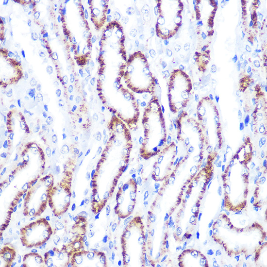
Immunohistochemistry analysis of paraffin-embedded Rat kidney using Smad4 Rabbit pAb (A5657) at dilution of 1:100 (40x lens). Microwave antigen retrieval performed with 0.01M PBS Buffer (pH 7.2) prior to IHC staining. |
|
|
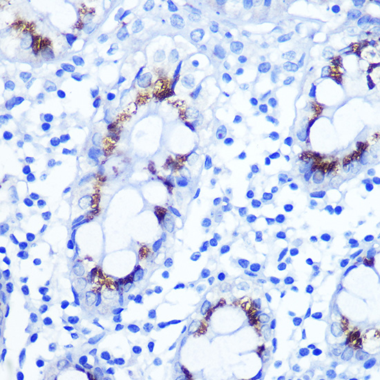
Immunohistochemistry analysis of paraffin-embedded Human colon using Smad4 Rabbit pAb (A5657) at dilution of 1:100 (40x lens). Microwave antigen retrieval performed with 0.01M PBS Buffer (pH 7.2) prior to IHC staining. |
|
|
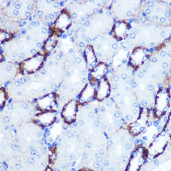
Immunohistochemistry analysis of paraffin-embedded Mouse kidney using Smad4 Rabbit pAb (A5657) at dilution of 1:100 (40x lens). Microwave antigen retrieval performed with 0.01M PBS Buffer (pH 7.2) prior to IHC staining. |
|
|
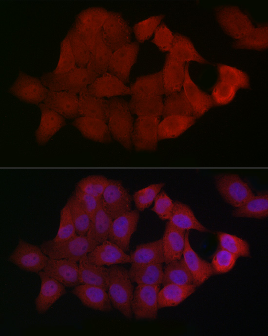
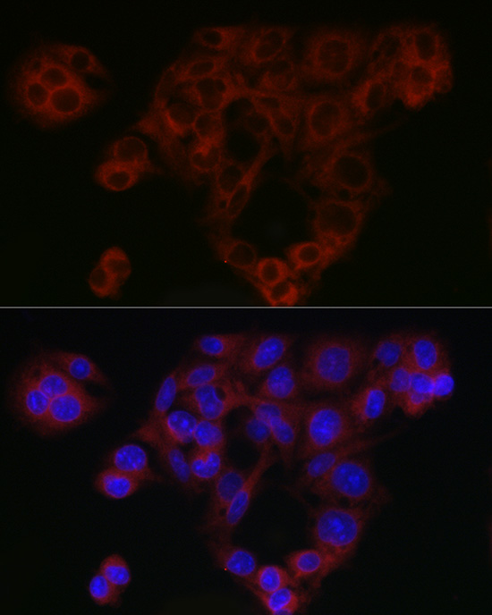
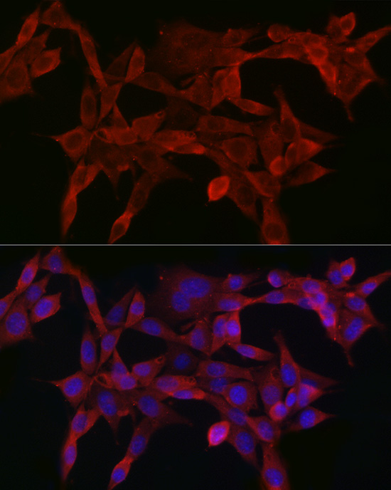
Immunofluorescence analysis of C6 cells using Smad4 Rabbit pAb (A5657) at dilution of 1:50 (40x lens). Secondary antibody: Cy3-conjugated Goat anti-Rabbit IgG (H+L) (AS007) at 1:500 dilution. Blue: DAPI for nuclear staining. |
|
|
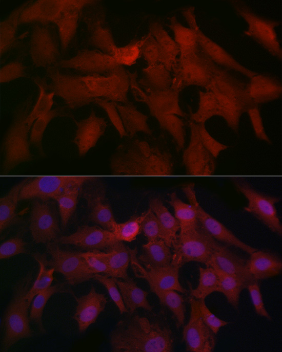
Immunofluorescence analysis of HeLa cells using Smad4 Rabbit pAb (A5657) at dilution of 1:50 (40x lens). Secondary antibody: Cy3-conjugated Goat anti-Rabbit IgG (H+L) (AS007) at 1:500 dilution. Blue: DAPI for nuclear staining. |
|
|
Immunofluorescence analysis of HepG2 cells using Smad4 Rabbit pAb (A5657) at dilution of 1:50 (40x lens). Secondary antibody: Cy3-conjugated Goat anti-Rabbit IgG (H+L) (AS007) at 1:500 dilution. Blue: DAPI for nuclear staining. |
|
|
Immunofluorescence analysis of NIH/3T3 cells using Smad4 Rabbit pAb (A5657) at dilution of 1:50 (40x lens). Secondary antibody: Cy3-conjugated Goat anti-Rabbit IgG (H+L) (AS007) at 1:500 dilution. Blue: DAPI for nuclear staining. |
Product Guarantee and Expert Support
