
CD13/ANPEP Rabbit pAb, Unconjugated, Polyclonal
Catalog Number:
ABB-A5662
- Images (8)
| Article Name: | CD13/ANPEP Rabbit pAb, Unconjugated, Polyclonal |
| Biozol Catalog Number: | ABB-A5662 |
| Supplier Catalog Number: | A5662 |
| Alternative Catalog Number: | ABB-A5662-100UL,ABB-A5662-20UL,ABB-A5662-1000UL,ABB-A5662-500UL |
| Manufacturer: | ABclonal |
| Host: | Rabbit |
| Category: | Antikörper |
| Application: | ELISA, IF, IHC-P, WB |
| Species Reactivity: | Human |
| Immunogen: | Recombinant protein (or fragment).This information is considered to be commercially sensitive. |
| Conjugation: | Unconjugated |
| Alternative Names: | APN, AP-M, AP-N, CD13, LAP1, P150, PEPN, hAPN, GP150, CD13/ANPEP |
| Aminopeptidase N is located in the small-intestinal and renal microvillar membrane, and also in other plasma membranes. In the small intestine aminopeptidase N plays a role in the final digestion of peptides generated from hydrolysis of proteins by gastric and pancreatic proteases. Its function in proximal tubular epithelial cells and other cell types is less clear. The large extracellular carboxyterminal domain contains a pentapeptide consensus sequence characteristic of members of the zinc-binding metalloproteinase superfamily. Sequence comparisons with known enzymes of this class showed that CD13 and aminopeptidase N are identical. The latter enzyme was thought to be involved in the metabolism of regulatory peptides by diverse cell types, including small intestinal and renal tubular epithelial cells, macrophages, granulocytes, and synaptic membranes from the CNS. This membrane-bound zinc metalloprotease is known to serve as a receptor for the HCoV-229E alphacoronavirus as well as other non-human coronaviruses. This gene has also been shown to promote angiogenesis, tumor growth, and metastasis and defects in this gene are associated with various types of leukemia and lymphoma. |
| Clonality: | Polyclonal |
| Molecular Weight: | 110kDa |
| NCBI: | 290 |
| UniProt: | P15144 |
| Purity: | Affinity purification |
| Sequence: | LASAHKVPVTLALNNTLFLIEERQYMPWEAALSSLSYFKLMFDRSEVYGPMKNYLKKQVTPLFIHFRNNTNNWREIPENLMDQYSEVNAISTACSNGVPECEEMVSGLFKQWMENPNNNPIHPNLRSTVYCNAIAQGGEEEWDFAWEQFRNATLVNEADKLRAALACSKELWILNRYLSYTLNPDLIRKQDATSTIISITNNVIGQGLVWDFVQSNWKKLFNDYGGGSFSFSNLIQAVTRRFSTEYELQQLEQFK |
| Target: | ANPEP |
| Antibody Type: | Primary Antibody |
| Application Dilute: | WB,1:500 - 1:1000|IHC-P,1:50 - 1:200|IF/ICC,1:50 - 1:100|ELISA,Recommended starting concentration is 1 µg/mL. Please optimize the concentration based on your specific assay requirements. |
| Application Notes: | Cross-Reactivity: Human,Mouse,Rat. ResearchArea: Immunology Inflammation,CDs,Stem Cells,Hematopoietic Progenitors,Mesenchymal Stem Cells,Cardiovascular,Angiogenesis. Shipping: Ice Bag |
|
|
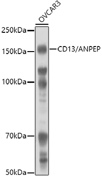
Western blot analysis of lysates from OVCAR3 cells, using CD13/ANPEP Rabbit pAb (A5662) at 1:1000 dilution. Secondary antibody: HRP-conjugated Goat anti-Rabbit IgG (H+L) (AS014) at 1:10000 dilution. Lysates/proteins: 25µg per lane. Blocking buffer: 3% nonfat dry milk in TBST. Detection: ECL Basic Kit (RM00020). Exposure time: 180s. |
|
|
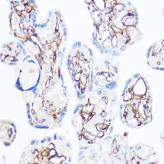
Immunohistochemistry analysis of paraffin-embedded Human placenta using CD13/ANPEP Rabbit pAb (A5662) at dilution of 1:50 (40x lens). High pressure antigen retrieval performed with 0.01M Citrate buffer (pH 6.0) prior to IHC staining. |
|
|
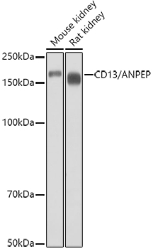
Western blot analysis of various lysates using CD13/ANPEP Rabbit pAb (A5662) at 1:1000 dilution. Secondary antibody: HRP-conjugated Goat anti-Rabbit IgG (H+L) (AS014) at 1:10000 dilution. Lysates/proteins: 25µg per lane. Blocking buffer: 3% nonfat dry milk in TBST. Detection: ECL Basic Kit (RM00020). Exposure time: 10s. |
|
|
Immunohistochemistry analysis of paraffin-embedded Mouse intestin using CD13/ANPEP Rabbit pAb (A5662) at dilution of 1:50 (40x lens). High pressure antigen retrieval performed with 0.01M Citrate buffer (pH 6.0) prior to IHC staining. |
|
|
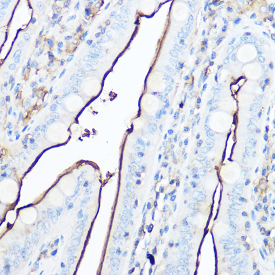
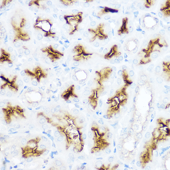
Immunohistochemistry analysis of paraffin-embedded Rat kidney using CD13/ANPEP Rabbit pAb (A5662) at dilution of 1:50 (40x lens). High pressure antigen retrieval performed with 0.01M Citrate buffer (pH 6.0) prior to IHC staining. |
|
|
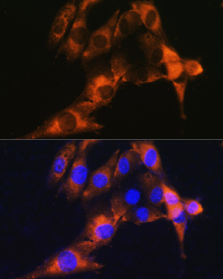
Immunofluorescence analysis of NIH/3T3 cells using CD13/CD13/ANPEP Rabbit pAb (A5662) at dilution of 1:100. Secondary antibody: Cy3-conjugated Goat anti-Rabbit IgG (H+L) (AS007) at 1:500 dilution. Blue: DAPI for nuclear staining. |
|
|
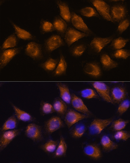
Immunofluorescence analysis of HeLa cells using CD13/CD13/ANPEP Rabbit pAb (A5662) at dilution of 1:100. Secondary antibody: Cy3-conjugated Goat anti-Rabbit IgG (H+L) (AS007) at 1:500 dilution. Blue: DAPI for nuclear staining. |
|
|
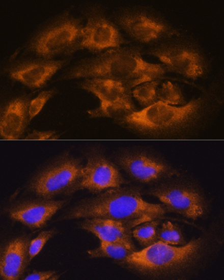
Immunofluorescence analysis of C6 cells using CD13/CD13/ANPEP Rabbit pAb (A5662) at dilution of 1:100. Secondary antibody: Cy3-conjugated Goat anti-Rabbit IgG (H+L) (AS007) at 1:500 dilution. Blue: DAPI for nuclear staining. |
Product Guarantee and Expert Support
