
PKC delta Rabbit mAb, Unconjugated, Monoclonal
Catalog Number:
ABB-A7778
- Images (8)

| Article Name: | PKC delta Rabbit mAb, Unconjugated, Monoclonal |
| Biozol Catalog Number: | ABB-A7778 |
| Supplier Catalog Number: | A7778 |
| Alternative Catalog Number: | ABB-A7778-100UL,ABB-A7778-20UL |
| Manufacturer: | ABclonal |
| Host: | Rabbit |
| Category: | Antikörper |
| Application: | ELISA, IF, IHC-P, WB |
| Species Reactivity: | Human |
| Immunogen: | Recombinant protein (or fragment).This information is considered to be commercially sensitive. |
| Conjugation: | Unconjugated |
| Alternative Names: | MAY1, PKCD, ALPS3, CVID9, nPKC-delta, PKC delta |
| The protein encoded by this gene is a member of the protein kinase C family of serine- and threonine-specific protein kinases. The encoded protein is activated by diacylglycerol and is both a tumor suppressor and a positive regulator of cell cycle progression. Also, this protein can positively or negatively regulate apoptosis. Defects in this gene are a cause of autoimmune lymphoproliferative syndrome. |
| Application Dilute: | WB,1:500 - 1:2000|IHC-P,1:50 - 1:200|IF/ICC,1:50 - 1:200|ELISA,Recommended starting concentration is 1 µg/mL. Please optimize the concentration based on your specific assay requirements. |
| Application Notes: | Cross-Reactivity: Human,Mouse,Rat. ResearchArea: Protein phosphorylation,Cancer,Signal Transduction,G protein signaling,G-Protein-Coupled Receptors Signaling to MAPK Erk,Kinase,Serine threonine kinases,ErbB-HER Signaling Pathway,MAPK-Erk Signaling Pathway,Cell Biology Developmental Biology,Apoptosis,Mitochondrial Control of Apoptosis,Inhibition of Apoptosis,TGF-b-Smad Signaling Pathway,Immunology Inflammation,B Cell Receptor Signaling Pathway,Neuroscience. Shipping: Ice Bag |
|
|
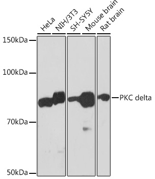
Western blot analysis of various lysates using PKC delta Rabbit mAb (A7778) at 1:1000 dilution incubated at room temperature for 1.5 hours. Secondary antibody: HRP-conjugated Goat anti-Rabbit IgG (H+L) (AS014) at 1:10000 dilution. Lysates/proteins: 25 µg per lane. Blocking buffer: 3% nonfat dry milk in TBST. Detection: ECL Basic Kit (RM00020). Exposure time: 5 s. |
|
|
Western blot analysis of lysates from C6 cells using PKC delta Rabbit mAb (A7778) at 1:1000 dilution incubated at room temperature for 1.5 hours. Secondary antibody: HRP-conjugated Goat anti-Rabbit IgG (H+L) (AS014) at 1:10000 dilution. Lysates/proteins: 25 µg per lane. Blocking buffer: 3% nonfat dry milk in TBST. Detection: ECL Basic Kit (RM00020). Exposure time: 45 s. |
|
|
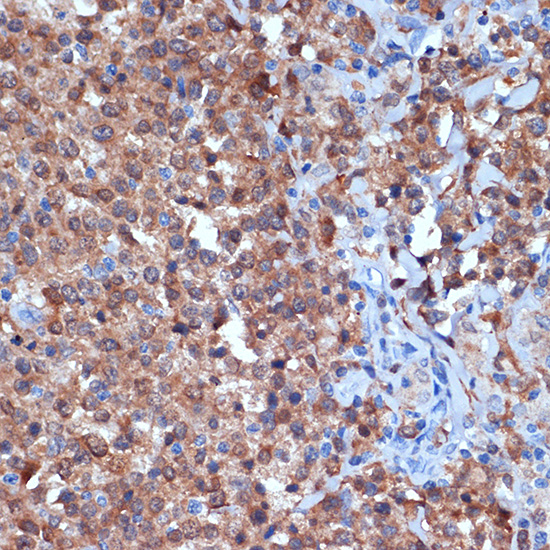
Immunohistochemistry analysis of paraffin-embeddedHuman spleen tissue usingPKC delta Rabbit mAb(A7778) at a dilution of 1:200 (40x lens).High pressure antigen retrieval was performed with 0.01 M citrate buffer (pH 6.0) prior to IHC staining. |
|
|
Immunohistochemistry analysis of paraffin-embeddedMouse colon tissue usingPKC delta Rabbit mAb(A7778) at a dilution of 1:200 (40x lens).High pressure antigen retrieval was performed with 0.01 M citrate buffer (pH 6.0) prior to IHC staining. |
|
|
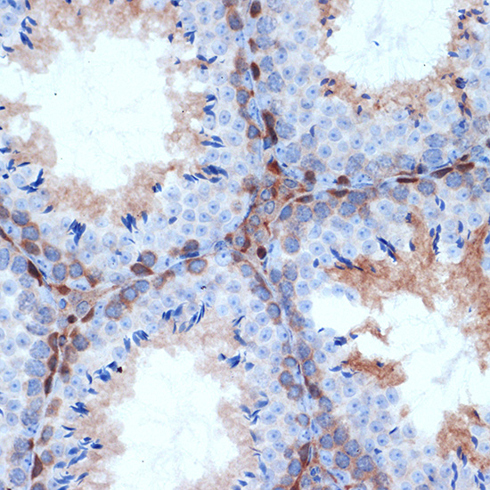
Immunohistochemistry analysis of paraffin-embeddedHuman thyroid tissue usingPKC delta Rabbit mAb(A7778) at a dilution of 1:200 (40x lens).High pressure antigen retrieval was performed with 0.01 M citrate buffer (pH 6.0) prior to IHC staining. |
|
|
Immunohistochemistry analysis of paraffin-embeddedMouse brain tissue usingPKC delta Rabbit mAb(A7778) at a dilution of 1:200 (40x lens).High pressure antigen retrieval was performed with 0.01 M citrate buffer (pH 6.0) prior to IHC staining. |
|
|
Immunohistochemistry analysis of paraffin-embeddedMouse testis tissue usingPKC delta Rabbit mAb(A7778) at a dilution of 1:200 (40x lens).High pressure antigen retrieval was performed with 0.01 M citrate buffer (pH 6.0) prior to IHC staining. |
|
|
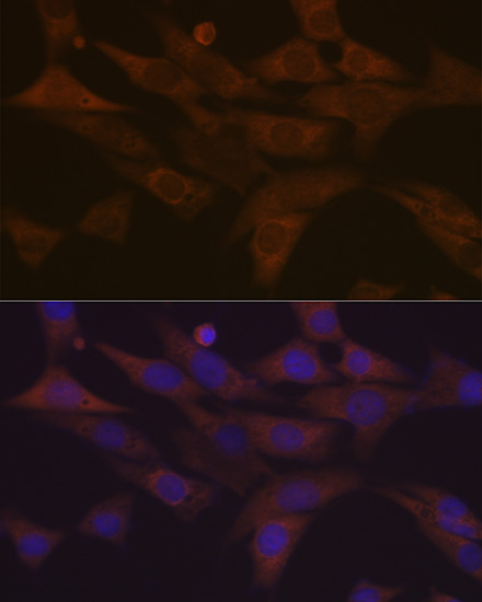
Immunofluorescence analysis of NIH-3T3 cells using PKC delta Rabbit mAb (A7778) at dilution of 1:100 (40x lens). Secondary antibody: Cy3-conjugated Goat anti-Rabbit IgG (H+L) (AS007) at 1:500 dilution. Blue: DAPI for nuclear staining. |
Product Guarantee and Expert Support
