
IL11RA Rabbit mAb, Unconjugated, Monoclonal
Catalog Number:
ABB-A9365
- Images (8)
| Article Name: | IL11RA Rabbit mAb, Unconjugated, Monoclonal |
| Biozol Catalog Number: | ABB-A9365 |
| Supplier Catalog Number: | A9365 |
| Alternative Catalog Number: | ABB-A9365-100UL,ABB-A9365-20UL |
| Manufacturer: | ABclonal |
| Host: | Rabbit |
| Category: | Antikörper |
| Application: | ELISA, IF, IHC-P, WB |
| Species Reactivity: | Human |
| Immunogen: | Synthetic peptide. This information is considered to be commercially sensitive. |
| Conjugation: | Unconjugated |
| Alternative Names: | CRSDA, IL11RA |
| Interleukin 11 is a stromal cell-derived cytokine that belongs to a family of pleiotropic and redundant cytokines that use the gp130 transducing subunit in their high affinity receptors. This gene encodes the IL-11 receptor, which is a member of the hematopoietic cytokine receptor family. This particular receptor is very similar to ciliary neurotrophic factor, since both contain an extracellular region with a 2-domain structure composed of an immunoglobulin-like domain and a cytokine receptor-like domain. Multiple alternatively spliced transcript variants have been found for this gene. |
| Application Dilute: | WB,1:500 - 1:1000|IHC-P,1:100 - 1:500|IF/ICC,1:50 - 1:200|ELISA,Recommended starting concentration is 1 µg/mL. Please optimize the concentration based on your specific assay requirements. |
| Application Notes: | Cross-Reactivity: Human,Mouse,Rat. ResearchArea: Cancer,Tumor biomarkers,Immunology Inflammation,Cytokines,Interleukins,Cell Intrinsic Innate Immunity Signaling Pathway,Stem Cells,Hematopoietic Progenitors. Shipping: Ice Bag |
|
|
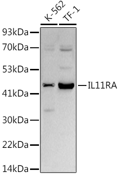
Western blot analysis of various lysates using (A9365) at 1:1000 dilution. Secondary antibody: HRP-conjugated Goat anti-Rabbit IgG (H+L) (AS014) at 1:10000 dilution. Lysates/proteins: 25µg per lane. Blocking buffer: 3% nonfat dry milk in TBST. Detection: ECL Basic Kit (RM00020). Exposure time: 30s. |
|
|
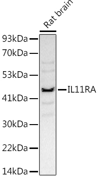
Western blot analysis of lysates from Rat brain, using (A9365) at 1:1000 dilution. Secondary antibody: HRP-conjugated Goat anti-Rabbit IgG (H+L) (AS014) at 1:10000 dilution. Lysates/proteins: 25µg per lane. Blocking buffer: 3% nonfat dry milk in TBST. Detection: ECL Basic Kit (RM00020). Exposure time: 180s. |
|
|
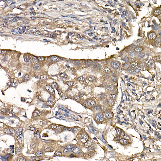
Immunohistochemistry analysis of paraffin-embedded Human colon tissue using IL11RA Rabbit mAb (A9365) at a dilution of 1:400 (40x lens). High pressure antigen retrieval performed with 0.01M Citrate buffer (pH 6.0) prior to IHC staining. |
|
|
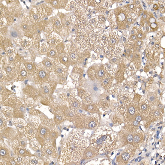
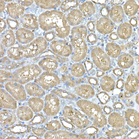
Immunohistochemistry analysis of paraffin-embedded Human liver cancer tissue using IL11RA Rabbit mAb (A9365) at a dilution of 1:400 (40x lens). High pressure antigen retrieval performed with 0.01M Citrate buffer (pH 6.0) prior to IHC staining. |
|
|
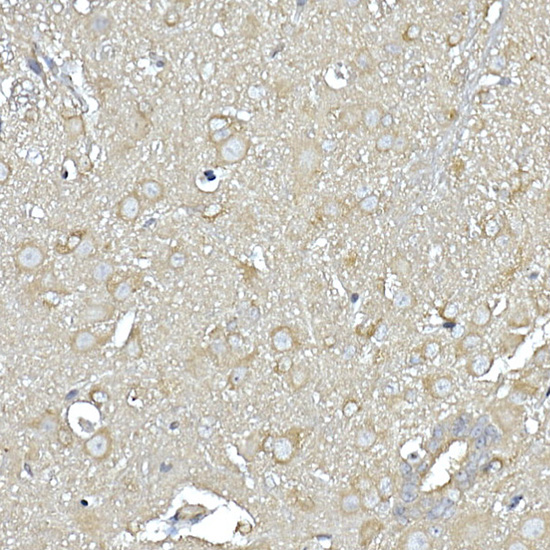
Immunohistochemistry analysis of paraffin-embedded Mouse brain tissue using IL11RA Rabbit mAb (A9365) at a dilution of 1:400 (40x lens). High pressure antigen retrieval performed with 0.01M Citrate buffer (pH 6.0) prior to IHC staining. |
|
|
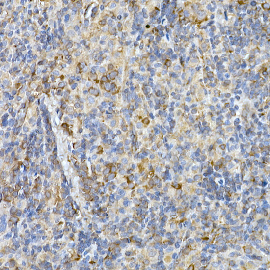
Immunohistochemistry analysis of paraffin-embedded Rat spleen tissue using IL11RA Rabbit mAb (A9365) at a dilution of 1:400 (40x lens). High pressure antigen retrieval performed with 0.01M Citrate buffer (pH 6.0) prior to IHC staining. |
|
|
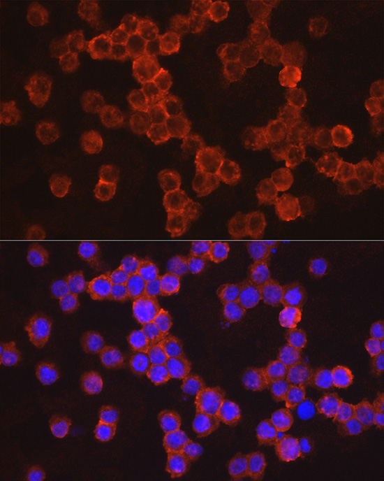
Confocal imaging of RAW 264.7 cells usingIL11RA Rabbit mAb (A9365,dilution 1:100) followed by a further incubation with Cy3 Goat Anti-Rabbit IgG (H+L) (AS007, dilution 1:500) (Red). DAPI was used for nuclear staining (Blue). Objective: 100x. |
|
|
Confocal imaging of K-562 cells usingIL11RA Rabbit mAb (A9365, dilution 1:100) followed by a further incubation with Cy3 Goat Anti-Rabbit IgG (H+L) (AS007, dilution 1:500)(Red). DAPI was used for nuclear staining (Blue). Objective: 100x. |
Product Guarantee and Expert Support
